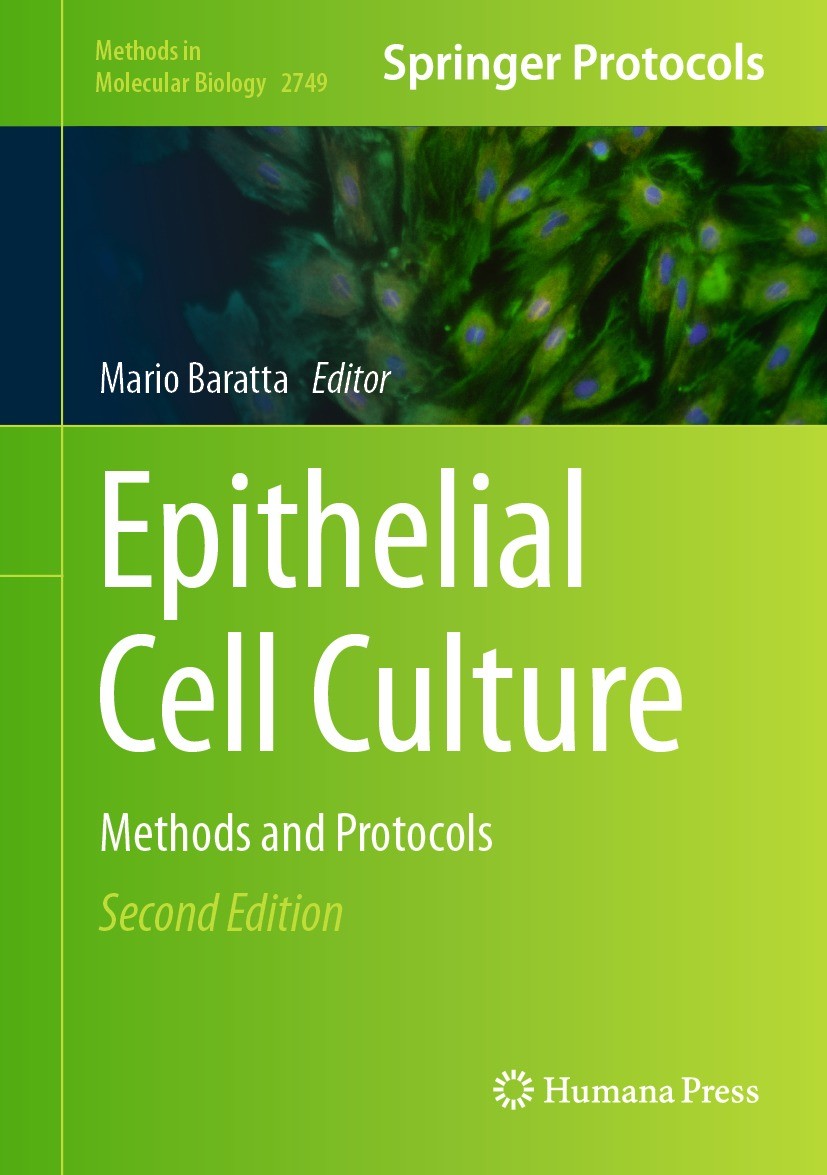
书目名称 | Epithelial Cell Culture | 副标题 | Methods and Protocol | 编辑 | Mario Baratta | 视频video | http://file.papertrans.cn/314/313372/313372.mp4 | 概述 | Includes cutting-edge methods and protocols.Provides step-by-step detail essential for reproducible results.Contains key notes and implementation advice from the experts | 丛书名称 | Methods in Molecular Biology | 图书封面 |  | 描述 | .Back Cover Copy..This second edition volume expands on the previous edition with in-depth discussions on the rapid advancements in epithelial cell biology, and the cutting-edge research and techniques used by researchers in the field. The chapters in this book cover topics such as detailed methodologies applicable to epithelial cells derived from primates, pigs, bovines, and laboratory animals; the manipulation and differentiation of epithelial cells; and epithelial cell models in the gastroenteric system in human medicine and nutrition. Written in the highly successful .Methods in Molecular Biology. .series format, chapters include introductions to their respective topics, lists of the necessary materials and reagents, step-by-step, readily reproducible laboratory protocols, and tips on troubleshooting and avoiding known pitfalls..Comprehensive and cutting-edge, .Epithelial Cell Culture: Methods and Protocols, Second Edition. .is a valuable resource for researchers in the scientific community, educators,and students who are interested in unraveling the complexities of epithelial cell biology, cultivating curiosity, and inspiring the next generation of groundbreaking research... . | 出版日期 | Book 2024Latest edition | 关键词 | gastrointestinal stem cells; intestinal organoids; Tumor Microenvironment; drug permeability; Zebrafish | 版次 | 2 | doi | https://doi.org/10.1007/978-1-0716-3609-1 | isbn_softcover | 978-1-0716-3611-4 | isbn_ebook | 978-1-0716-3609-1Series ISSN 1064-3745 Series E-ISSN 1940-6029 | issn_series | 1064-3745 | copyright | The Editor(s) (if applicable) and The Author(s), under exclusive license to Springer Science+Busines |
1 |
Front Matter |
|
|
Abstract
|
2 |
,Culture of Mouse Thymic Epithelial Cells in Serum-Free Medium, |
Yasuhiro Adachi |
|
Abstract
Primary cell culture systems are widely used as a valuable method for analyzing the biological functions of specific cells in vitro. Recently, various serum-free primary cell culture methods have been developed that do not involve the use of animal serums. Since the thymus is comprised of many cell types, such as thymocytes, thymic epithelial cells, macrophages, and fibroblasts, thymic epithelial cells must be isolated for their functional analysis in vitro. This chapter describes the detailed protocol for the selective primary culture of thymic epithelial cells using defined serum-free medium.
|
3 |
,Cultured Pig Thyroid Follicular Cells: Electrical Evaluation of Epithelial Integrity, |
Peying Fong |
|
Abstract
Thyroid epithelial cells organize as enclosed follicles containing thyroid hormone precursor, iodinated thyroglobulin, with lumina bordered by the cellular apices. Transepithelial transport determines composition of compartmental milieu essential for both prohormone formation and its downstream conversion to thyroxine. Hence, not only do follicular lumina function as storage vessels but also as physiological reaction chambers into which reactive components, together with the proper salts and water, are secreted. Polarized, two-dimensional cultures of pig thyroid epithelia, prepared using established protocols, provide a convenient system for assessing transport processes subserving hormone production. This chapter details established methods for growing and evaluating integrity of primary pig thyroid cultures for downstream analysis of transport and other key physiological functions.
|
4 |
,Human Minor Salivary Glands: A Readily Available Source of Salivary Stem/Progenitor Cells for Regen |
Caitlynn M. L. Barrows,Danielle Wu,Simon Young,Mary C. Farach-Carson |
|
Abstract
Resident stem/progenitor cells within the secretory salivary glands offer a potential therapeutic resource for use in the regeneration of salivary glands needed to restore saliva production in patients with chronic xerostomia, or dry mouth. Methods were developed previously to isolate human stem/progenitor cells (hS/PCs) from major salivary glands (parotid/submandibular). Abundant minor salivary glands located in readily accessible locations in the oral cavity and lip could provide an additional valuable therapeutic resource. An advantage of this cell resource is that these minor glands about the size of grape seeds can be harvested from healthy donors using minimally invasive surgical procedures. The disadvantage of using minor glands is that they contain many fewer cells than do major glands, and thus harvested cells need to be expanded in the lab to create a therapeutic resource. While earlier work has described isolation of proliferative cell populations from minor salivary glands that could be used in regenerative medicine, most of these expanded cells possess properties of mesenchymal cells rather than the epithelial population that secretes salivary products..Here, we descri
|
5 |
,Salivary Organotypic Tissue Culture: An Ex-vivo 3D Model for Studying Radiation-Induced Injury of H |
Akshaya Upadhyay,Migmar Tsamchoe,Simon D. Tran |
|
Abstract
An organotypic tissue culture model can maintain the cellular and molecular interactions, as well as the extracellular components of a tissue ex vivo. Thus, this 3D model biologically mimics in vivo conditions better than commonly used 2D culture in vitro models. Here, we provide a detailed workflow for generating live 3D organotypic tissue slices from patient-derived freshly resected salivary glandular tissues. We also cover the processing of these tissues for various downstream applications like live-dead viability/cytotoxicity assay, FFPE sectioning and immunostaining, and RNA and protein extraction with a focus on the salivary gland radiation injury model. These procedures can be applied extensively to various solid organs and used for disease modeling for cancer research, radiation biology, and regenerative medicine.
|
6 |
,Differentiation of Pig Gastric Primary Cells into Mucus Producing Epithelial Cells, |
Macarena P. Quintana-Hayashi,Sinan Sharba,Sara K. Lindén |
|
Abstract
There is a growing interest in the development of in vitro models that mimic the intrinsic characteristics of cells in vivo to replace and/or reduce the use of experimental animals. The stomach is lined with mucus secreting epithelial cells, creating a thick mucus layer that protects the underlying epithelial cells from acid, pathogens, and other harmful agents. Mucins are a main component of the mucus layer, and their secretion is an important protective feature of epithelial cells in vivo. Here, we present a method that differentiates pig gastric primary cells into mucin secreting epithelial cells by culturing the cells on polyester membranes under semi-wet interface for 14 days, using differentiation medium containing the .-[(3,5-difluorophenyl)acetyl]-L-alanyl-2-phenyl]glycine-1,1-dimethylethyl ester (DAPT) in the basolateral compartment for the first 7 days and subsequent 7-day culture in non-differentiation medium. The in vitro mucosal surfaces created by these cells are harvested 2 weeks post confluence, and two preservation methods are described to fix the monolayers for further analysis.
|
7 |
,Isolation, Culture, and Microscopic Imaging of Guinea Pig Primary Gastric Tissue Cells, |
Weronika Gonciarz,Magdalena Chmiela |
|
Abstract
In this chapter, the procedure of isolation and propagation of guinea pig gastric tissue primary cells in cell culture in vitro is presented. Selected methods of microscopic imaging of cells are shown, including monitoring the ability of cells to migrate as a determinant of their activity. The primary cells that expanded in cell cultures in vitro have characteristics of natural cells and facilitate studying both the spontaneous and induced biological processes on the cellular level. Particularly, the primary cells derived from the guinea pig stomach were found to be a good model for studying the effects of bacteria-host interactions and the development of inflammatory responses driven by gastric pathogen ..
|
8 |
,Method for Two-Dimensional Epithelial Monolayer Formation Derived from Mouse Three-Dimensional Smal |
Yuta Takase,Toshio Takahashi |
|
Abstract
The intestinal epithelium is composed of two distinct structures, namely, the villi and crypts. The base of the crypts contains intestinal stem cells (ISCs), which support the high regenerative capacity of the intestinal epithelium. With the establishment of the three-dimensional (3D) organoid culture method, the cellular and molecular mechanisms of differentiation, proliferation, and maintenance of ISCs have been widely analyzed. However, the sphere-like morphology of the 3D organoids prevents access to the apical side of the epithelium. To overcome this limitation, two-dimensional (2D) monolayer cultures derived from 3D organoids have been attempted; however, 2D culture methods for the mouse small intestine have not been well established. In this study, we developed a simple method that uses only commercially available materials, for the formation of 2D epithelial monolayers from mouse 3D small intestinal organoids. Using this method, confluent 2D epithelial monolayers were established within 4 days. This monolayer showed stable tight junction and included ISCs and differentiated intestinal cells. It also showed physiologically relevant transepithelial electrical resistance value
|
9 |
,Human Hepatic Spheroid Coculture Model for the Assessment of Drug-Induced Liver Injury, |
Linhao Li,Hongbing Wang |
|
Abstract
Accurate evaluation of potential drug risks such as drug-induced liver injury (DILI) continues to be a challenge faced by pharmaceutical industry and regulatory agencies. Preclinical testing has served as a foundation for the evaluation of the potential risks and effectiveness of investigational new drug (IND) products in humans. However, current two-dimensional (2D) in vitro human primary hepatocyte (HPH) culture systems cannot accurately depict and simulate the rich environment and complex processes observed in vivo, while animal studies present inherited species-specific differences and low throughput scales. Thus, there is a continued demand to establish new approaches that can better characterize DILI during drug discovery and development. Among others, the three-dimensional (3D) hepatic spheroid model comprising self-aggregated primary human hepatocytes cocultured with non-parenchymal cells (NPCs) appears to be a more accurate representation of the natural hepatic microenvironment with intercellular interactions between hepatocytes, stellate cells, Kupffer cells, liver sinusoidal endothelial cells (LSECs), and other cell types. This model holds the potential to improve the ab
|
10 |
,In Vitro Porcine (Explant) Colon Culture, |
Matheus de Oliveira Costa,Michael K. Dame |
|
Abstract
Models have been extensively used to investigate disease pathogenesis. Animal models are costly and require extensive logistics for animal care, and samples are not always suitable for different analytical techniques or to answer the research question. In vitro cell culture models are generally focused on recreating a specific characteristic of an organ and are limited to a single cell population that does not display the characteristic tissue architecture of the source organ. In addition, such models do not account for the many interactions between pathogens and the diverse cell subsets that are normally present in a given organ. Conclusions based on conventional 2D cell culture methods are limited, requiring extrapolation from a reductionist model to understand in vivo events. In vitro organ culture (IVOC) offers a way to overcome some of these limitations. Explants conserve important in vivo characteristics, such as different cell types and complex tissue architecture. This in vitro (ex vivo) organ culture protocol of the swine large intestine aims at maintaining viable colonic mucosa for up to 5 days. The protocol described herein applies a combination of methods used for immor
|
11 |
,Bioencapsulation of Oocytes and Granulosa Cells, |
Massimo Faustini,Stella Agradi,Daniele Vigo,Maria L. Torre,Giulio Curone |
|
Abstract
A protocol for the encapsulation in sodium alginate of granulosa cells in primary culture and coculture of oocyte-cumulus complexes is reported. Sodium alginate forms strong gels when jellified with barium ions, allowing the self-organization of cells into a 3D structure. This method of encapsulation is simple and cheap, allowing the culture of cells in a three-dimensional fashion.
|
12 |
,Culturing and Differentiation of Patient-Derived Ectocervical Epithelial Stem Cells Using Air-Liqui |
Rajendra Kumar Gurumurthy,Naveen Kumar,Cindrilla Chumduri |
|
Abstract
The ectocervix acts as a multilayered defense barrier, protecting the female reproductive system from external pathogens and supporting fertility and pregnancy. To understand the complex cellular and molecular mechanisms of cervical biology and disease, reliable in vitro models are vital. We present an efficient method to isolate and cultivate epithelial stem cells from ectocervical tissue biopsies. This method combines enzymatic digestion, mechanical dissociation, and selective culturing to obtain pure ectocervical epithelial cells for further investigation. The protocol accommodates both 2D stem cell monolayer and advanced 3D culture systems, such as air-liquid interface and Matrigel scaffolds, using a defined media cocktail, making it highly versatile. The primary ectocervical epithelial cells retain their native characteristics, enabling the exploration of ectocervical epithelial tissue behavior and pathology. This chapter provides step-by-step guidelines for setting up 2D and 3D cultures, facilitating adoption across different laboratories, and advancing cervical biology and disease research.
|
13 |
,Ovine Trophoblast Cells: Cell Isolation and Culturing from the Placenta at the Early Stage of Pregn |
Paola Toschi,Irene Viola,Isabella Manenti,Silvia Miretti,Elisabetta Macchi,Eugenio Martignani,Paolo |
|
Abstract
Embryo development is dependent upon the exchange of oxygen and nutrients through the placenta, mainly composed of peculiar epithelioid cells, known as trophoblast cells. Normal trophoblast functionality plays a key role during the whole pregnancy, especially in the first stage of placentation. This chapter explains the techniques to obtain sheep primary trophoblast cells from the early placenta. Overall, procedures for cell isolation, culture, characterization, and cryopreservation are described.
|
14 |
,Amniotic Membrane and Amniotic Epithelial Cell Culture, |
Angelo Canciello,Adrian Cerverò-Varona,Maura Turriani,Valentina Russo,Barbara Barboni |
|
Abstract
Amniotic membrane (AM) is considered an important medical device for applications in regenerative medicine. The therapeutic properties of AM are due to its resistant extracellular matrix and to the large number of bioactive molecules released by its cells. To this regard, ovine amniotic epithelial cells (AECs) are a subset of placental stem cells with great regenerative and immunomodulatory properties. Indeed, either oAEC or AM have been object of intense study for regenerative medicine, thanks to several advantages in developing preclinical studies on a high value translational animal model, such as sheep. For this reason, a critical standardization of cultural practices is fundamental in order to maintain, on one hand, AM integrity and structure and, on the other hand, oAEC native properties, thus improving their in vivo therapeutic potential and clinical outcomes..In addition, freshly isolated AECs or AM can be exploited to produce enriched immunomodulatory secretomes that had been used with success into cell-free regenerative medicine procedures..To this aim, here is described an improved oAEC cultural protocol able to preserve their native epithelial phenotype also after the i
|
15 |
,Evaluation of the Epithelial Barrier Integrity in Primary Cultures of Pig Mammary Epithelial Cells, |
Chiara Bernardini,Debora La Mantia,Monica Forni |
|
Abstract
A major feature of epithelial and endothelial cells is the creation of biological barriers able to protect the body against stressors that could compromise homeostasis. The ability to characterize biological barriers in vitro is an important study tool especially used for the intestinal barrier, the blood-brain barrier, and the lung barrier. The strength and integrity of biological barriers may be assessed by the measurement of the transepithelial/transendothelial electrical resistance (TEER) that reflects the ionic conductance of the paracellular pathway. The TEER measurement is a quantitative, non-invasive, highly useful, and representative method that must be strictly standardized. Here we describe a quantitative protocol to assess the mammary epithelial barrier integrity by combining the TEER measurement with a test for studying the passage of the sodium fluorescein, that is, a hydrophilic paracellular marker. Being the swine species an excellent translational model, primary cultures of mammary epithelial cells, isolated from hybrid pig tissue collected at slaughterhouse, are used.
|
16 |
,Bovine Skeletal Muscle Satellite Cells: Isolation, Growth, and Differentiation, |
Silvia Miretti,Isabella Manenti,Paola Toschi,Elisabetta Macchi,Eugenio Martignani,Paolo Accornero,Ma |
|
Abstract
Skeletal muscle in cattle occupies a large part of the animal’s body mass and develops into an important source of nutrients for human nutrition. Recently, the attention on bovine myogenic cells is increased to develop strategies of cultured in vitro meat as an alternative food source, more sustainable, ethical, and healthy than traditional meat production. At present, investigating the proliferation and differentiation of bovine skeletal muscle myogenic cells in vitro maintains its importance in the study of the mechanisms underlying the physiological and pathological events affecting the skeletal muscle, but it is of particular interest in animal husbandry and the food industry fields..In cell-based biological research, cell lines are one of the favored experimental tools because a population of cells could proliferate indefinitely in vitro under different stimuli, but they are limited to addressing the relevant biological properties of a cell population. On the other hand, primary cells from normal animal tissues undergo a limited number of divisions in vitro before they enter senescence but preserve their original characteristics and functions, and researchers can acquire the o
|
17 |
,Equine Induced Pluripotent Stem Cell Culture, |
Julia Falk,F. Xavier Donadeu |
|
Abstract
Groundbreaking work by Takahashi and Yamanaka in 2006 demonstrated that non-embryonic cells can be reprogrammed into pluripotent stem cells (PSCs) by forcing the expression of a defined set of transcription factors in culture, thus overcoming ethical concerns linked to embryonic stem cells. Induced PSCs have since revolutionized biomedical research, holding tremendous potential also in other areas such as livestock production and wildlife conservation. iPSCs exhibit broad accessibility, having been derived from a multitude of cell types and species. Apart from humans, iPSCs hold particular medical promise in the horse. The potential of iPSCs has been shown in a variety of biomedical contexts in the horse. However, progress in generating therapeutically useful equine iPSCs has lagged behind that reported in humans, with the generation of footprint-free iPSCs using non-integrative reprogramming approaches having proven particularly challenging. A greater understanding of the underlying molecular pathways and essential factors required for the generation and maintenance of equine iPSCs and their differentiation into relevant lineages will be critical for realizing their significant po
|
18 |
Back Matter |
|
|
Abstract
|
|
|