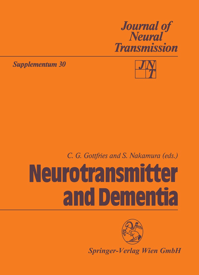
书目名称 | Neurotransmitter and Dementia | 编辑 | C. G. Gottfries,S. Nakamura | 视频video | | 丛书名称 | Journal of Neural Transmission. Supplementa | 图书封面 |  | 描述 | Neurotransmitter changes taking place in the brain of patients with dementia disorders, mainly Alzheimer type dementia, are reported. Their role in the pathogenesis of Alzheimer‘s disease is discussed; and the neurochemical changes are considered as a base for formulating treatment strategies. By studying markers in the cerebrospinal fluid, diagnostic methods may be achieved that will aid in diagnosing subgroups of dememtia. | 出版日期 | Conference proceedings 1990 | 关键词 | Alzheimer; CNS; alzheimer‘s disease; brain; dementia; metabolism; neurons; neurotransmitter; research | 版次 | 1 | doi | https://doi.org/10.1007/978-3-7091-3345-3 | isbn_softcover | 978-3-211-82190-9 | isbn_ebook | 978-3-7091-3345-3Series ISSN 0303-6995 | issn_series | 0303-6995 | copyright | Springer-Verlag Wien 1990 |
1 |
Front Matter |
|
|
Abstract
|
2 |
,Some effects of CNS cholinergic neurons on memory, |
T. Goto,F. Kuzuya,H. Endo,T. Tajima,H. Ikari |
|
Abstract
The aim of this study is to observe the relationship between the impairment in passive avoidance task induced in rats by the i. p. administration of muscarinic antagonists, scopolamine and methyl-scopolamine, and the change in acetylcholine (ACh) output induced by these drugs. Initially we studied the effects of these drugs on the animals’ performance of a step-through passive avoidance task. We then measured the change in ACh levels after administration of these drugs using an in vivo brain dialysis technique. Scopolamine was effective in impairing the performance of the passive avoidance task, while methyl-scopolamine did not have clear effects on the performance of the task. With regard to ACh output, scopolamine increased ACh dose-dependently and methyl-scopolamine also affected ACh release. These data suggest that the accumulation of ACh in the synaptic cleft may be involved in the memory deficit induced by scopolamine.
|
3 |
,Subcellular distribution of acetylcholinesterase in Alzheimer’s disease: abnormal localization and |
S. Nakamura,S. Kawashima,S. Nakano,T. Tsuji,W. Araki |
|
Abstract
AChE activity was detected mainly in membrane-bound fractions in the frontal cortex of autopsied control or Alzheimer brain as well as rat cerebral cortex. However, the distribution of AChE among various membrane fractions was different between control and Alzheimer brains. The highest specific activity was detected in the fraction enriched with senile plaque, which was obtained from the Alzheimer brain by sonication, solubilization with detergent and centrifugation on a sucrose density gradient. The senile plaque enriched fraction was incubated with purified collagenase or protease and centrifuged at 100,000 . for 60 min. More than 50% of AChE activity was detected in the supernatant fraction. AChE in the supernatant solution showed a property of G4 isozyme. AChE might probably be anchored to the senile plaque through its collagen tail and be solubilized with collagenase or protease, producing a G4 isozyme.
|
4 |
,Changes of acetylcholine and choline concentrations in cerebrospinal fluids of normal subjects and |
Y. Ikeda,S. Okuyama,Y. Fujiki,K. Tomoda,K. Ohshiro,T. Itoh,T. Yamauchi |
|
Abstract
Acetylcholine (ACh) and choline (Ch) in cerebrospinal fluid from 29 normal volunteers and 7 patients with Alzheimer-type dementia (DAT) were examined using high-performance liquid chromatography with electrochemical detector coupled with liquid cation-exchange method. In normal volunteers, ACh concentration was decreased significantly from 40–50 years and Ch concentration was increased significantly from 50–60 years. CSF from patients with DAT revealed high Ch concentration and the increase was statistically significant while ACh concentration in CSF of DAT did not show a significant difference with that of normal volunteers. This Ch augmentation may suggest a disturbance in utilization of Ch for ACh synthesis and may become an useful indicator for organic changes in central cholinergic system.
|
5 |
,Disturbance of the 5-hydroxytryptamine metabolism in brains from patients with Alzheimer’s dementia |
C. G. Gottfries |
|
Abstract
The 5-hydroxytryptamine (5-HT) system in the human brain is sensitive to aging. In dementia of the Alzheimer type (AD/SDAT), there are significantly reduced concentrations of 5-HT and 5-hydroxyindoleacetic acid (5-HIAA). 5-HT-sensitive imipramine binding is reduced by almost 50%, indicating a loss of presynaptic 5-HT terminals. There also seems to be reduced tryptophan hydroxylase activity in some brain areas. In cerebrospinal fluid (CSF) from AD/SDAT patients, the concentration of 5-HIAA is reduced, and the accumulation of 5-HIAA after probenecid loading is diminished. Biochemical findings together with structural findings in the raphe nuclei indicate that the disturbance of the 5-HT system is of the same magnitude as the disturbance of the cholinergic system..Reduced activity in the 5-HT system may be of importance for activity in the hypothalamus. There is an increased concentration of arginine vasopressin, which may explain the increased activity in the hypothalamic-pituitary-adrenal axis seen in patients with AD/SDAT. This activity is reduced when a selective 5-HT reuptake blocker is given..Pharmacological treatment with 5-HT reuptake blockers improves emotional disturbances,
|
6 |
,CSF ,-endorphin, HVA and 5-HIAA of dementia of the Alzheimer type and Binswanger’s disease in the e |
S. Lee M.D.,T. Chiba,T. Kitahama,R. Kaieda,M. Hagiwara,A. Nagazumi,A. Terashi |
|
Abstract
Cerebrospinal fluid (CSF) concentration of .-endorphin (.- Ep), homovanillic acid (HVA) and 5-hydroxyindoleacetic acid (5-HIAA) was measured in 15 patients with dementia of the Alzheimer type (DAT) and in 16 patients suspected of having Binswanger’s disease (BD) by MRI, which sometimes resembles DAT clinically. These were classified into three stages according to severity of dementia, Stage 1 (mild dementia)-Stage 3 (severe dementia). CSF levels of HVA decreased significantly in severe dementia, but the level of 5-HIAA did not correlate with dementia severity in both dementia groups. .-Ep levels did not differ significantly between any stages of DAT, and among controls. .-Ep levels, however, in BD Stage 1 (27.5 ± 5.9 pg/ml) were significantly higher (p < 0.05), but level in Stage 3 (6.7 ± 2.0) was significantly lower (p < 0.001) than in the controls (19.2 ± 4.5). These results suggest that CSF .-Ep may depend on the cause of dementia rather than severity of dementia, and could possibly distinguish the closely resembling BD from true DAT.
|
7 |
,Neuropeptide levels in Alzheimer’s disease and dementia with frontotemporal degeneration, |
L. Minthon M.D.,L. Edvinsson,R. Ekman,L. Gustafson |
|
Abstract
The CSF levels of somatostatin-LI (SLI), neuropeptide Y (NPY-LI) and Delta Sleep Inducing Peptide (DSIP-LI) have been measured in patients with dementia of Alzheimer type (DAT) and dementia with frontotemporal degeneration of non-Alzheimer type (FTD). The distribution pattern of cortical degeneration differs between these two types of dementia. DAT shows degeneration of mainly temporo-parietal and temporo-limbic structures, whereas FTD discloses its main degeneration in the frontotemporal regions (Brun, 1987). The somatostatin-LI was significantly reduced both in DAT and FTD. NPY-LI showed a significant reduction in DAT but not in FTD. A tendency to a reduction with duration of the disease was observed in DAT whereas the contrary was noted in FTD. The DSIP-LI levels were reduced in DAT and slightly increased in FTD. The study provides an evidence of neurochemical differences between the two primary degenerative dementias.
|
8 |
,Changes in signal transduction in Alzheimer’s disease, |
S. Shimohama,H. Ninomiya,T. Saitoh,R. D. Terry,R. Fukunaga,T. Taniguchi,M. Fujiwara,J. Kimura,M. Kam |
|
Abstract
We studied the signal transduction system including the receptor and protein kinase C (PKC) in Alzheimer’s disease (AD) brains. We used .H-TCP as a ligand for the NMDA receptor-ion channel complex. The total concentrations of .H-TCP binding sites were significantly reduced in AD frontal cortex. .H-TCP binding sites spared in AD brains retained the affinity for the ligand and the reactivity to NMDA, .-glutamate, and glycine. We utilized antibodies to assess the degree of involvement of different PKC isoforms in AD. The concentration of PKC (.II) was lower in AD particulate fractions and higher in AD cytosol fractions. Immunocytochemical studies revealed reduced numbers of anti-PKC (.II)-immunopositive neurons. Anti-PKC (.) faintly stained entire plaques and surrounding glial cells. Anti-PKC (.I) stained dystrophic plaque neurites. Anti-PKC (.II) stained the amyloid-containing portions of plaques. These results suggest an involvement of second messenger cascades in the pathogenesis of AD in addition to neurotransmitters and their receptors.
|
9 |
Back Matter |
|
|
Abstract
|
|
|