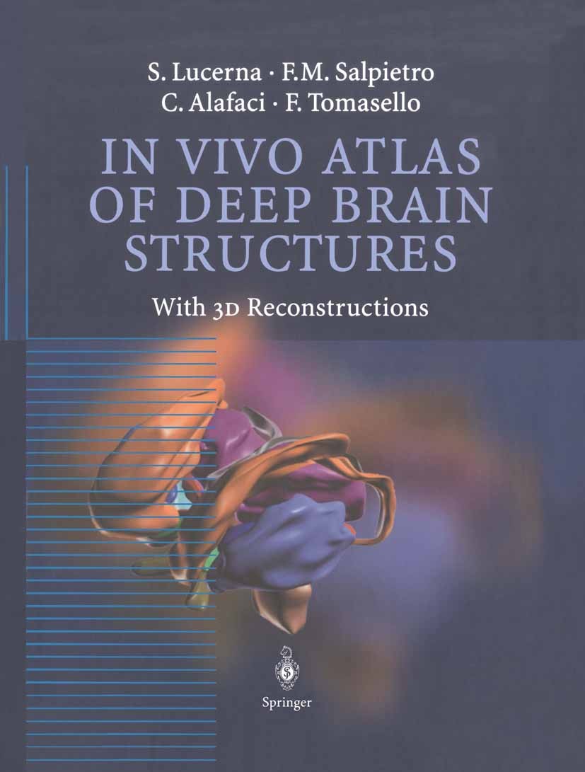
| 书目名称 | In Vivo Atlas of Deep Brain Structures | | 副标题 | With 3D Reconstructi | | 编辑 | Sebastiano Lucerna,Francesco M. Salpietro,Francesc | | 视频video | http://file.papertrans.cn/464/463041/463041.mp4 | | 概述 | Useful millimetric scale and highest possible precision of the images make the atlas an ideal tool for surgical planning.Superb quality of 4-color 2D and 3D reconstructions | | 图书封面 |  | | 描述 | In the first half of the twentieth century, the study of neuroanatomy was essentiallybased on the observations made by scientists on brain cadavers fixed with standard techniques. These studies have produced well-known tools such as the stereotactic atlas, which have proven to be extremely useful and irreplaceable for neurosurgeons, neuroradi ologists, neurologists and neuroanatomists. In particular, the Talairach and Schaltenbrandt atlases are considered the most presti gious and up-to-date work available today. The recent introduction of neuroimaging, especially nuclear magnetic resonance, together with the exciting and tremendous progress made in computer graphics, has allowed us to approach neuroanatomy directly in living patients with more accuracy and a high degree ofdetail. This work, after a short introduction which explains the methodolo gy used, is divided into four types of sections: three types ofsections obtained from the same brain and orientated in the standard axial, sagittal, and coronal spatial planes and one type of section of three dimensional pictures obtained from the computerized processing of the previous pictures. The organization and the life-size tabl | | 出版日期 | Book 2002 | | 关键词 | MRI atlas; brain; magnetic resonance; magnetic resonance imaging (MRI); neuroscience; three dimensional r | | 版次 | 1 | | doi | https://doi.org/10.1007/978-3-642-56381-2 | | isbn_softcover | 978-3-642-62710-1 | | isbn_ebook | 978-3-642-56381-2 | | copyright | Springer-Verlag Berlin Heidelberg 2002 |
The information of publication is updating

|
|
 |Archiver|手机版|小黑屋|
派博传思国际
( 京公网安备110108008328)
GMT+8, 2026-2-9 21:17


