找回密码
 To register

QQ登录

只需一步,快速开始

扫一扫,访问微社区

Titlebook: Diagnostic Imaging in Ophthalmology; Carlos F. Gonzalez (Professor of Radiology),Melvin Book 1986 Springer-Verlag New York, Inc. 1986 Tumo

[复制链接]
查看: 53969|回复: 56
发表于 2025-3-21 19:12:23 | 显示全部楼层 |阅读模式
书目名称Diagnostic Imaging in Ophthalmology
编辑Carlos F. Gonzalez (Professor of Radiology),Melvin
视频video
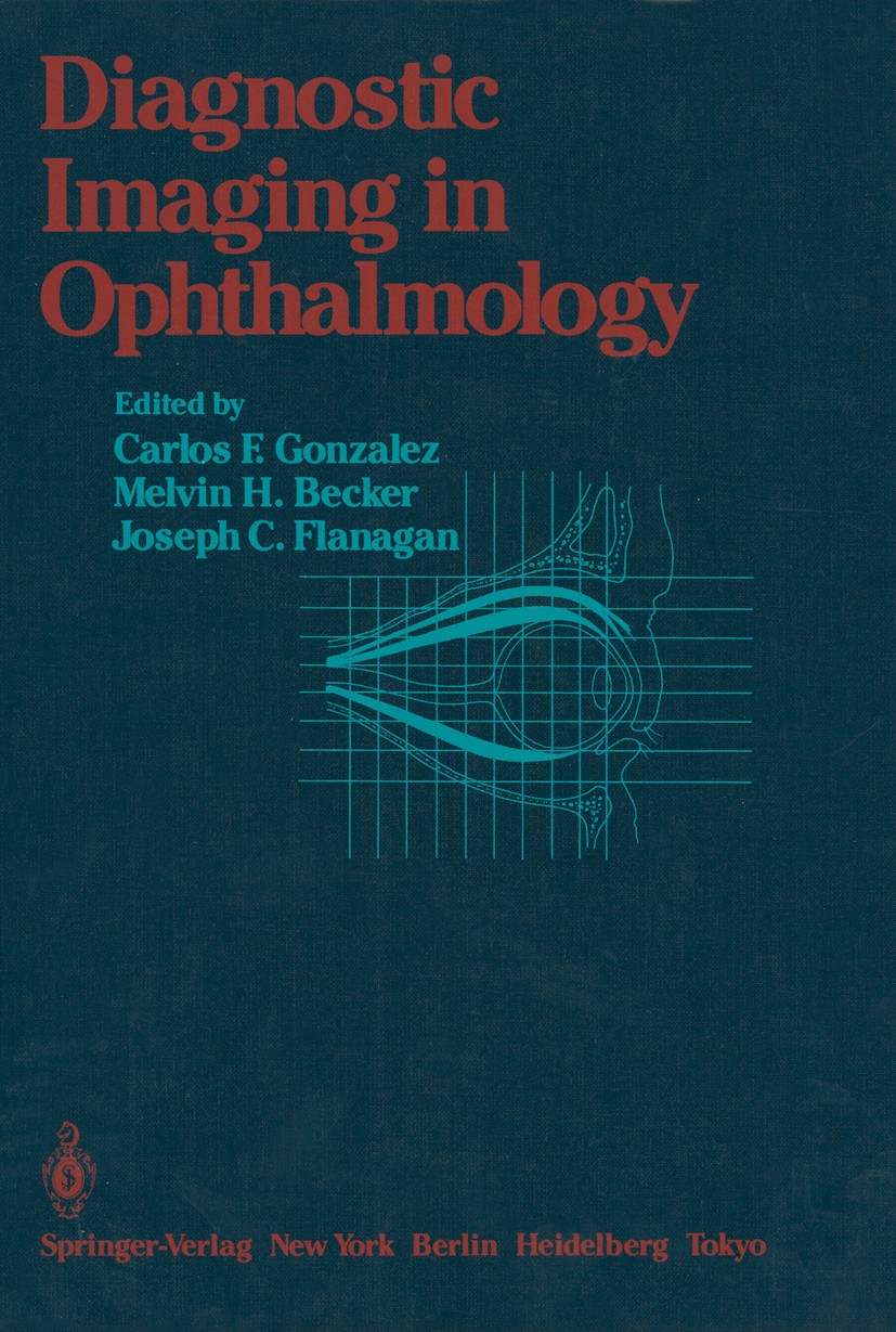
图书封面Titlebook: Diagnostic Imaging in Ophthalmology;  Carlos F. Gonzalez (Professor of Radiology),Melvin Book 1986 Springer-Verlag New York, Inc. 1986 Tumo
描述This book has been written for radiologists, ophthalmologists, neurologists, neurosur­ geons, plastic surgeons, and others interested in the evaluation of disorders with ophthalmologic signs and symptoms. It is designed to provide recent knowledge in this area derived from ultrasonography, computed tomography (CT), and magnetic resonance imaging (MRI). In the past decade, the advent of ultrasonography, computed tomography, and more recently magnetic resonance imaging has provided diagnostic images of the eye, orbit, and brain in a fashion that had been a dream of many prior to the develop­ ment of these techniques. These newer modes of diagnosis have replaced some previous techniques, such as nuclear medicine imaging and, to some degree, vascular studies and orbitography. There are three sections to this book. The first section is a discussion of the imaging techniques. The second is devoted to the role of these imaging methods in the evaluation of ophthalmic disorders. The last section, dealing with radiotherapy for ophthalmologic tumors, is included because the current imaging techniques are needed for treatment planning. We wish to thank the many people who have assisted us in p
出版日期Book 1986
关键词Tumor; brain; computed tomography (CT); diagnosis; diagnostic imaging; eye; infection; magnetic resonance; m
版次1
doihttps://doi.org/10.1007/978-1-4613-8575-2
isbn_softcover978-1-4613-8577-6
isbn_ebook978-1-4613-8575-2
copyrightSpringer-Verlag New York, Inc. 1986
The information of publication is updating

书目名称Diagnostic Imaging in Ophthalmology影响因子(影响力)


书目名称Diagnostic Imaging in Ophthalmology影响因子(影响力)学科排名


书目名称Diagnostic Imaging in Ophthalmology网络公开度


书目名称Diagnostic Imaging in Ophthalmology网络公开度学科排名


书目名称Diagnostic Imaging in Ophthalmology被引频次


书目名称Diagnostic Imaging in Ophthalmology被引频次学科排名


书目名称Diagnostic Imaging in Ophthalmology年度引用


书目名称Diagnostic Imaging in Ophthalmology年度引用学科排名


书目名称Diagnostic Imaging in Ophthalmology读者反馈


书目名称Diagnostic Imaging in Ophthalmology读者反馈学科排名


单选投票, 共有 1 人参与投票
 

0票 0.00%

Perfect with Aesthetics


0票 0.00%

Better Implies Difficulty


0票 0.00%

Good and Satisfactory


0票 0.00%

Adverse Performance


1票 100.00%

Disdainful Garbage

您所在的用户组没有投票权限
发表于 2025-3-21 22:44:10 | 显示全部楼层
发表于 2025-3-22 03:16:01 | 显示全部楼层
Investigation of the Orbit by Contrast Techniques,evelopment of highly sophisticated computed tomographic (CT) and ultrasound techniques has decreased their use. Vascular studies are used only in the investigation of vascular malformations or vascular tumors within or around the orbit. Contrast injections into the orbital soft tissues (contrast orb
发表于 2025-3-22 05:37:15 | 显示全部楼层
The Lacrimal Drainage System,of these is epiphora, which is the abnormal overflow of tears and usually is secondary to obstruction of the passages. Other conditions that require study are diverticula, fistulae, masses in the area of or within the lumen of the lacrimal passages, persistent drainage or obstruction after surgery o
发表于 2025-3-22 09:01:21 | 显示全部楼层
发表于 2025-3-22 14:24:15 | 显示全部楼层
发表于 2025-3-22 18:48:42 | 显示全部楼层
Congenital Abnormalities,). The conditions that involve the bony structures are the ones in which imaging techniques—such as routine radiographs, tomograms, and computed tomography (CT)—are of most value. A knowledge of both the embryologie development and anatomic measurements of the orbits is helpful in understanding the
发表于 2025-3-22 23:46:40 | 显示全部楼层
Orbital Tumors,nents. Secondary tumors may involve the orbit from adjacent structures, such as the paranasal sinuses, the eyeball, the intracranial structures, and the eyelids; or they may be metastatic lesions from distant structures. (Table 11.1). Inflammations, pseudotumors, and vascular lesions also appear as
发表于 2025-3-23 05:10:31 | 显示全部楼层
发表于 2025-3-23 06:36:10 | 显示全部楼层
 关于派博传思  派博传思旗下网站  友情链接
派博传思介绍 公司地理位置 论文服务流程 影响因子官网 SITEMAP 大讲堂 北京大学 Oxford Uni. Harvard Uni.
发展历史沿革 期刊点评 投稿经验总结 SCIENCEGARD IMPACTFACTOR 派博系数 清华大学 Yale Uni. Stanford Uni.
|Archiver|手机版|小黑屋| 派博传思国际 ( 京公网安备110108008328) GMT+8, 2025-5-20 14:02
Copyright © 2001-2015 派博传思   京公网安备110108008328 版权所有 All rights reserved
快速回复 返回顶部 返回列表