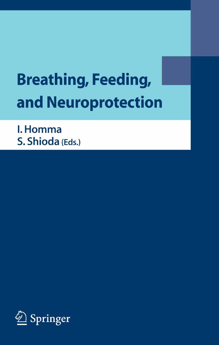
| 期刊全称 | Breathing, Feeding, and Neuroprotection | | 影响因子2023 | Ikuo Homma (Professor and Chairman),Seiji Shioda ( | | 视频video | http://file.papertrans.cn/191/190644/190644.mp4 | | 发行地址 | Prior works in respiratory pattern generation | | 图书封面 |  | | 影响因子 | .New findings in brain research are being revealed on an almost daily basis, and the focus of this book is the fields of breathing, neuroprotection, and higher brain functions. An unresolved issue within respiration research and hence a topic of much interest is Where and how respiratory rhythm is generated in the brainstem, detailed analysis of which is presented herein. Chapters on neuroprotection examine the functional significance of the blood – brain barrier as an interface of blood and the central nervous system; other chapters look at health and disease in relation to the hypothalamic and limbic systems. In addition to animal experiments, research on the human brain is included, with a focus on the recently developed EEG/dipole tracing method. This book will be an invaluable reference for researchers in neuroscience and related fields.. | | Pindex | Conference proceedings 2006 |
| 1 |
Front Matter |
|
|
Abstract
|
| 2 |
|
|
|
Abstract
|
| 3 |
Visualization of Respiratory Neuron Activity in the Ventral Medulla from a Newborn Rodent |
Hiroshi Onimaru,Akiko Arata,Satoru Arata,Ikuo Homma |
|
Abstract
We visualized respiratory neuron network activity in the medulla of the rat and mouse in vitro by optical recordings using voltage-sensitive dye. The brainstem and spinal cord of 0- to 1-day-old Wistar rats and 0-day-old mice isolated under deep ether anaesthesia were incubated in a modified Krebs solution containing a fluorescent voltage-sensitive dye. Fluorescence signals corresponding to respiratory activity were detected by a CCD image sensor. Pre-inspiratory neuron activity appeared in the limited region of the rostral ventrolateral medulla [i.e. para-facial respiratory group (pFRG) region], preceding the onset of inspiratory activity by about 500 ms. During the inspiratory phase, plateau activity appeared in the more caudal ventrolateral medulla at the level of most rostral roots of the XIIth nerve (i.e. the pre-Bötzinger complex level). We found that pre-inspiratory neurons which were a predominant subtype of the pFRG neurons were located in the area immediately beneath the ventral pia mater at the level of the facial nucleus. We also analyzed respiratory neuron activity in the wild type and two kinds of knock-out mice that exhibit respiratory failure leading to neonatal dea
|
| 4 |
Search for Genes Involved Central Respiratory Failure Using Mice Models |
Satoru Arata,Hiroshi Onimaru,Akiko Arata,Ikuo Homma,Tomio Inoue,Seiji Shioda,Seiji Shirasawa,Michael |
|
Abstract
To search for genes that cause human congenital respiratory failure, we undertook several studies using wild type mice and gene-deficient mice that exhibit respiratory failure. 1) To assess respiratory abnormality, ventilatory parameters were determined by whole-body plethysmography of prenatal and neonatal mice. 2) To clarify the mechanism of respiratory failure, respiratory neuron activity was examined by electrophysiological and optical imaging analysis using a brainstem-spinal cord preparation. 3) To search for the genes related to central respiratory failure in brainstem, alteration of gene expression in the medulla oblongata during transition from embryonic respiration to external respiration was examined by DNA microarray. To date, we have studied several lines of gene-deficient mice exhibit respiratory failure, and we propose a model of central respiratory failure as a syndrome resembling congenital central hypoventilation.
|
| 5 |
Control of Central Histaminergic Neurons for Respiration during Hypercapnia in Conscious Mice |
Michiko Iwase,Masahiko Izumizaki,Kenichi Miyamoto,Mitsuko Kanamaru,Ikuo Homma |
|
Abstract
Central histaminergic neurons located in the posterior hypothalamus affect many autonomic behaviors. We examined the roles of these neurons in respiration in conscious histamine H1 receptor-knockout (H1RKO) and wild-type (WT) mice. Acute stepwise hypercapnia increased respiratory frequency (f), tidal volume (V.), and minute ventilation (V.) in both genotypes. However, H1RKO mice showed a lower f response and a higher VT response than those of WT mice. The V.-T. relation curve for H1RKO mice was shifted to the right and upward relative to that for WT mice, suggesting that in H1RKO mice the termination of inspiration is delayed by an increase in the inspiratory off switch (IOS) threshold. Increased body temperature increased the f response to hypercapnia in WT but not in H1RKO mice. This polypnea during hypercapnia was due mainly to a reduction in T.. These results suggest that central histaminergic neurons contribute to the termination of inspiration in the IOS mechanism and to polypnea during hyperthermia via activation of H1 receptors.
|
| 6 |
|
|
|
Abstract
|
| 7 |
The Role of the Blood-Brain Barrier in Feeding: Leptin |
William A. Banks,Susan A. Farr,John E. Morley |
|
Abstract
The blood- brain barrier (BBB) has emerged as a regulatory interface that controls the exchange of informational molecules between the blood and the central nervous system (CNS) fluids (the brain interstitial and the cerebrospinal fluid). As such, it is a pivotal point in a humoral-based, endocrine-like communication between the CNS and peripheral tissues. For example, the BBB controls the entry of major feeding hormones into the CNS from the blood, including leptin. Impaired transport of leptin across the BBB is an early cause of leptin resistance. Leptin transport is regulated by alpha.-adrenergics, which simulate transport, and triglycerides, which inhibit it. The ability of triglycerides to inhibit leptin transport may have evolved as a response to the hypertriglyceridemia of starvation. If so, their ability to inhibit leptin transport in obesity may be a metabolic case of mistaken identity.
|
| 8 |
Ultrastructural Study on the Demyelination of the White Matter in the Rat Spinal Cord after Cardiac |
Li Yin,Yoshifumi Kudo,Hirokazu Ohtaki,Sachiko Yofu,Masaji Matsunaga,Seiji Shioda |
|
Abstract
Neuronal damages in lumbar spinal cord are known to induce the motor deficit after systemic ischemia including cardiac arrest and heart attack. Recently we reported that the neuronal damages in the hippocampus and lumbar spinal cord were induced after the combination of cardiac arrest and massive hemorrhagic shock. In the model, the neurons in intermediate gray matter were showed necrotic neuronal cell death at 1 d after ischemia. However, the damages of white matter did not understood well. Therefore, we focused on the white matter damages and have checked on the processes of the demyelination in limber spinal cord after severe systemic ischemia using electron microscopy. At 1 d, the myelin sheaths was collapsed and dissociated from axon although the axons seemed relatively intact. At 7 d, many shrank axons were observed and some of axons were disappeared. This result suggested the progressively demyelination was observed in ventral white matter in lumber spinal cord prior to axonal degeneration after severe systemic ischemia.
|
| 9 |
Engagement of Proinflammatory Cytokines after Cerebral Ischemia |
Hirokazu Ohtaki,Li Yin,Kenji Dohi,Sachiko Yofu,Tomoya Nakamachi,Masaji Matsunaga,Seiji Shioda |
|
Abstract
The incidence of stroke is gradually increasing in the industrialized world and a major cause of long-lasting disability. To determine the mechanism of neuronal cell death after stroke and develop the new strategy of therapy, several studies had been performed during the past decade and some proinflammatory cytokines were identified as the therapeutic candidate. However, the critical actions of proinflammatory cytokines are still largely unknown in detail because the cytokines play a role on small amount, show the short half-life, and have multiple functions for depending on the amount and time of expression. We present here the pathophysiological events of cerebral ischemia and the action and possibility of proinflammatory cytokines from the recent results.
|
| 10 |
Levels of the Alkoxy Radical in Patients with Brain Death |
Kenji Dohi,Kazue Satoh,Hiroshi Moriwaki,Yuko Mihara,Yasufumi Miyake,Hirokazu Ohtaki,Seiji Shioda,Toh |
|
Abstract
Lipid peroxidation is induced by reactive oxygen species and is involved in acute neuronal damage. Thus, controlling this process may be a realistic therapeutic strategy for treating or preventing neuronal damage. However, the study of free radicals in body fluids is currently severely hampered by technical difficulties in their detection. We have developed an ex vivo electron spin resonance (ESR) method, that employs 5,5-dimethyl-1-pyrroline-N-oxide (DMPO) as a spin trap, to measure the alkoxyl radical (OR) in human blood. We found that this method can detect OR produced by treating human blood ex vivo. Analysis of jugular venous and radial arterial blood from patients with acute neuronal damage revealed higher OR levels in the venous blood compared to arterial samples. In brain death patients, however, the jugular vein OR spectrum was similar to the radial artery OR level. The ratio between radial artery and jugular vein levels, expressed as the RI (radical intensity) ratio = aORI/jORI, was calculated. A rterial-jugular bulb RI ratio below 1, together with accepted clinical criteria (unresponsive coma with brainstem areflexia), may provide a non-invasive assessment of cerebral ci
|
| 11 |
Differentiation of Neural Stem Cells into Astrocytes by Low Concentration of Pituitary Adenylate Cyc |
Jun Watanabe,Seiji Shioda,Sakae Kikuyama,Kazuyasu Nakaya,Shigeo Nakajo |
|
Abstract
Recently it has been reported that pituitary adenylate cyclase-activating polypeptide (PACAP) possesses diverse physiological functions in central nervous system. In this study, the effect of physiological concentrations of PACAP on the differentiation of neural stem cells is described. When neural stem cells were exposed to 0.2 nM PACAP, total number of the cells decreased during the 8 days-culture. However, number of MAP2-immunopositive cells decreased while GFAP-immunopositive cells increased depending on the concentration of PACAP (0.2–2 nM). Neural stem cells were differentiated into astrocytes even at a concentration below 2 nM. These results indicate that PACAP at a low concentration prefers induction of differentiation into astrocytes rather than proliferation in neural stem cells.
|
| 12 |
Neurogenesis is Toxicologically Heterogeneous: A Case of BrdU-lnduced Cell Death |
Tetsuo Ogawa,Makiko Kuwagata,Seiji Shioda |
|
Abstract
In developmental neuroscience, 5-bromo-2′-deoxyuridine (BrdU) is extensively used as a useful tool for labeling proliferating cells. Doses ranging from 10 to 100 mg/kg are commonly injected into animals. However, several lines of evidence demonstrated that BrdU has genotoxicity. The present study demonstrates that 1) a single injection of BrdU at 100 mg/kg into pregnant mice on gestation day 11.5 induced cell death in the neuroepithelium of certain brain areas such as the frontal neocortex, but not other areas such as the mesencephalic tegmentum and pons, 2) injections of BrdU into adult mice at 100 or 300 mg/kg did not induce cell death in the subventricular zone (SVZ) of the lateral ventricle, known to be a site of adult neurogenesis. These findings strongly suggest that 1) sensitivity to the genotoxicity differes among fetal neural stem cells, 2) BrdU-labeling should be avoided when early neurogenesis is being observed.
|
| 13 |
|
|
|
Abstract
|
| 14 |
Hypocretin-Deficient Narcolepsy as a Disease Model to Study the Hypothalamic Function in Health and |
Seiji Nishino |
|
Abstract
Using forward (i.e. positional cloning in canine narcolepsy) and reverse (i.e. mouse gene knockout) genetics, genes involved in the pathogenesis of narcolepsy (hypocretin/orexin ligand and its receptor) in animals have been identified. Mutations in hypocretin related-genes are rare in humans, but hypocretin-ligand deficiency is found in many cases. This discovery is likely to lead to the development of new diagnostic tests and treatments. Hypocretins/orexins are novel hypothalamic neuropetides also involved in various hypothalamic functions such as energy homeostasis, autonomic and neuroendocrine functions. Hypocretin-deficient narcolepsy thus appears now to be a more complex condition than a simple sleep disorder, and it may serve a disease model to study the most fundamental hypothalamic function.
|
| 15 |
The Expression of Synaptic Vesicle Proteins after Chronic Antidepressant Treatment in Rat Brain |
Misa Yamada,Kou Takahashi,Chika Kurahashi,Mitsuhiko Yamada,Kazuo Honda |
|
Abstract
The biological basis for the therapeutic mechanisms of depression are still unknown. We have previously performed EST analysis and identified some common biological changes induced after chronic antidepressant treatment as antidepressant related genes/ESTs: ADRG#1-707. Then, we developed our original cDNA microarray on which ADRG#1-707 were spotted, for rapid secondary screening of candidate genes as the novel therapeutic targets. With this microarray, we found that the expression of some of the ADRGs were related to neurotransmiter release and located on synaptic vesicle. Indeed, VAMP2/synaptobrevin, cysteine string protein, synapsin I, Rab-IA and Rab-3B were induced after chronic sertraline treatment in rat frontal cortex. Western blot analysis also demonstrated the induction of these ADRGs at protein levels after chronic treatment with imipramine and sertraline. In addition, synaptophysin and secretogranin II, often used as a marker protein for small synaptic vesicle or large dense core granule were significantly increased after chronically treatment with antidepressants. On the other hand, the expression of SNAP-25 and syntaxin-1, which are used as markers for synapse and make
|
| 16 |
Identification of Molecular Systems Responsible for the Therapeutic Effect of Antidepressant |
Mitsuhiko Yamada,Misa Yamada |
|
Abstract
Although blockade by antidepressants of monoamine uptake into nerve endings is one of the cornerstones of the monoamine hypothesis of depression, there is a clear discrepancy between the rapid effects of antidepressants in increasing synaptic concentrations of monoamine and the lack of immediate clinical efficiency of antidepressant treatment. Pharmacogenomics, functional genomics and proteomics are powerful tools that can be used to identify genes/ESTs or molecular systems affected by antidepressants. Using a differential cloning strategy, we and other groups have isolated genes that are differentially expressed in the brain after chronic antidepressant treatment. Some of these candidate genes may encode functional molecular systems or pathways induced by chronic antidepressant treatment. Defining the roles of these molecular systems in drug-induced neural plasticity is likely to transform the course of research on the biological basis of depression. Such detailed knowledge will have profound effects on the diagnosis, prevention, and treatment of depression.
|
| 17 |
S-100B Expression in Neonatal Rat Cortical “Barrels” and Thalamic “Barreloids” |
Katsumasa T. Muneoka,Hisayuki Funahashi,Tetsuo Ogawa,Makiko Kuwagata,Patricia M. Whitaker-Azmitia,Se |
|
Abstract
“Barreloids” and “barrels” are patch-like structure representing single whiskers observed in the ventroposterior thalamic nucleus (VP) and somatosensory cortex in rodents, respectively. They are characteristic structure observed during the early neonatal period, which is an important period for the development of the networks in VP and somatosensory cortex. Various neurotransmitter systems have been reported to be involved in the development of these structure including, serotonergic, glutamatargic, GABAergic and cholinergic ones. The present study indicated that immunoreactivity for a calcium binding protein, S-100B, was transiently found as “barreloids” in VP at postnatal day (PND) 7 and as “barrels” in the somatosensory layer IV at PND 15, respectively. “Barrel” - like 5-hydroxytriptamine transporter (5-HTT) immunoreactivity was also found in the somatosensory cortex at PND 15. Morphological findings indicated that S-100B was present in cellular nuclei and released into extracellular space in both the thalamus and cortex. S-100B is suggested to be involved in the formation or remodeling of networks in VP and the somatosensory cortex via modulating calcium levels intra- and extra
|
| 18 |
Postnatal Change of Glycinergic Synaptic Transmission from Supratrigeminal Region to Trigeminal Moto |
Tomio Inoue,Shiro Nakamura,Kan Nakajima,Kotaro Maki |
|
Abstract
We have investigated excitatory synaptic transmission from the lateral part of the supratrigeminal region (lSuV) to the trigeminal motor nucleus (MoV) in neonatal and juvenile rat brain stem slice preparations by high-speed optical recording techniques and gramicidin perforated-patch recordings. Electrical stimulation of lSuV evoked optical responses in MoV. An antidromic response in lSuV was evoked by MoV stimulation while synaptic transmission was suppressed by substitution of external Ca. with Mn.. Application of CNQX and APV to MoV reduced the optical responses in MoV evoked by ISuV stimulation in both neonatal and juvenile rats. Application of strychnine to MoV also suppressed the optical responses in MoV of neonatal rats. On the other hand, strychnine enhanced the optical responses in MoV of juveniles. Gramicidin perforated-patch recordings from trigeminal motoneurons (TMNs) revealed that glycinergic postsynaptic potentials evoked by lSuV stimulation were depolarizing in neonatal rats but become hyperpolarizing in juveniles. We conclude that inputs from lSuV excite TMNs through activation of glutamate or glycine receptors in neonatal rats, whereas glycine receptor activation
|
| 19 |
Central Nervous System Concerned with the Stress Induced Inhibition of Cellular Immune Activity |
Takao Sato,Shiyu Guo,Tadashi Hisamitsu |
|
Abstract
Many studies about the effect of stressors on immune activities were reported, but the mechanisms of those effects were obscure yet. Restraint water immersion stress reduced splenic NK cell activity (NK activity) measured by cytotoxicity to YAC-1 cells in rats through both humoral and neural mechanisms. Electrical lesion of hypothalamic paraventricular nucleus (PVN) markedly enhanced NK activity, but the lesion of ventromedial hypothalamic nucleus (VMH) depressed NK activity. These results suggest that the stress may inhibit NK activity through the activation of hypothalamic paraventricular nucleus.
|
| 20 |
|
|
|
Abstract
|
书目名称Breathing, Feeding, and Neuroprotection影响因子(影响力) 
书目名称Breathing, Feeding, and Neuroprotection影响因子(影响力)学科排名 
书目名称Breathing, Feeding, and Neuroprotection网络公开度 
书目名称Breathing, Feeding, and Neuroprotection网络公开度学科排名 
书目名称Breathing, Feeding, and Neuroprotection被引频次 
书目名称Breathing, Feeding, and Neuroprotection被引频次学科排名 
书目名称Breathing, Feeding, and Neuroprotection年度引用 
书目名称Breathing, Feeding, and Neuroprotection年度引用学科排名 
书目名称Breathing, Feeding, and Neuroprotection读者反馈 
书目名称Breathing, Feeding, and Neuroprotection读者反馈学科排名 
|
|
|