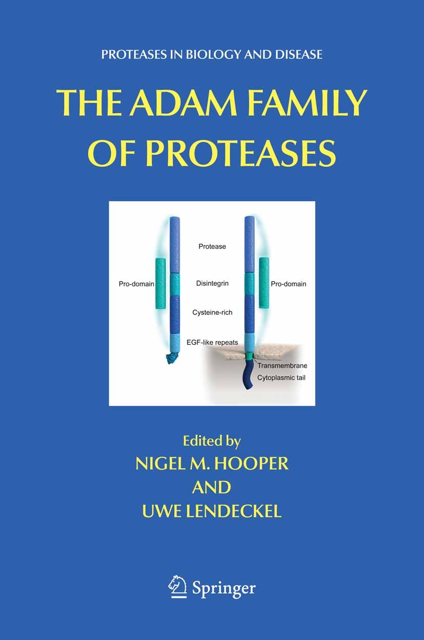
书目名称 | The ADAM Family of Proteases | 编辑 | Nigel M. Hooper,Uwe Lendeckel | 视频video | | 概述 | The ADAM Family of Proteases will be a timely and useful source of information both for those immersed in the field and those just entering this exciting and rapidly expanding field of research. The i | 丛书名称 | Proteases in Biology and Disease | 图书封面 |  | 描述 | .The ADAM Family of Proteases provides the first comprehensive review of the roles of ADAMs and the related ADAMTS proteases in biology and disease. Although a few members of the ADAM (a disintegrin and metalloprotease) family have been known for some time, it is only in recent years through advances in genome sequencing that the large size of this family of zinc metalloproteases has become apparent. These proteins have multiple domains including a protease domain and a disintegrin domain. A branch of the family, called ADAMTS, also have thrombospondin-like motifs. The role of ADAMs and ADAMTS members in a diversity of biological processes is gradually coming to light. For example, some ADAMs have critical roles in the ectodomain shedding of membrane proteins including tumour necrosis factor-a, the cell signalling molecule Notch and the Alzheimer’s amyloid precursor protein. Other ADAM and ADAMTS family members have key roles to play in sperm function and fertility, collagen processing, development, cardiac hypertrophy and arthritis. . | 出版日期 | Book 2005 | 关键词 | biology; genome; protein; proteins; sequencing | 版次 | 1 | doi | https://doi.org/10.1007/b106833 | isbn_softcover | 978-1-4419-3775-9 | isbn_ebook | 978-0-387-25151-6 | copyright | Springer-Verlag US 2005 |
1 |
,Introduction to the ADAM Family, |
Judith White,Lance Bridges,Douglas DeSimone,Monika Tomczuk,Tyra Wolfsberg |
|
Abstract
ADAMs (proteins containing A Disintegrin and A Metalloprotease domain) are multidomain and multifunctional proteins that are emerging as key regulators of critical events that occur at the cell surface. Many ADAMs (roughly half) are active metalloproteases, and several of these (e.g. ADAMs 10, 17, and 19) exert important functions ., for example in development of the heart and brain. The best-characterized . activity of ADAM proteases is as ectodomain sheddases. By shedding cell surface proteins (e.g. cytokines and growth factors), ADAMs initiate extracellular signaling events (e.g. signaling through epidermal growth factor receptors). ADAM-mediated ectodomain shedding (e.g. of Notch) can also set the stage for important intracellular signaling events. ADAMs have also been reported to shed surface proteins involved in both cell-cell and cell-matrix adhesion. The disintegrin and cysteine-rich domains of ADAMs exhibit adhesive activities in tissue culture-based studies. The important roles that several proteolytically inactive ADAMs play in development (ADAMs 2, 3, 14, and 23) suggest that ADAM adhesive activities may be relevant to their function. In this chapter, we first review the history and phylogeny of the ADAMs as well as structural and functional aspects of their major domains. We next review how ADAMs function as ectodomain sheddases, how their protease activities may be regulated, and how ADAMs may function in modulating cell adhesion and cell migration. We end with a very brief discussion of the role of ADAMs in development and disease and conclude by posing some questions for future research. Our goal is to give an appreciation for the widespread, varied, and fascinating means by which ADAMs affect, or may affect, key cell surface events: cell signaling, cell adhesion, and cell migration.
|
1 |
,Studies from ADAM Knockout Mice, |
Keisuke Hoiruchi,Carl P. Blobel |
|
Abstract
ADAMs are membrane anchored glycoproteins that contain a disintegrin and metalloprotease domain. This chapter will focus on recent insights that have emerged from studies of “knockout” mice for ADAM proteases that are widely expressed or at least expressed in a variety of different cells and tissues (ADAMs 8, 9, 10, 12, 15, 17 and 19). These studies have shown that ADAM10 is important for signaling via the cell surface receptor Notch during development, while ADAM17 is critical for the development of the lung, epithelial structures and semilunar heart valves because of its role in the functional activation of ligands of the epidermal growth factor receptor. ADAM19 is essential for proper development of heart valves and the ventricular septum, although the underlying mechanism remains to be established. On the other hand, ADAMs 8, 9, 12 and 15 are dispensable for normal development and adult life in mice, at least under laboratory conditions. However, ADAM15 has a critical role in pathological neovascularization, making it a potential target for the design of inhibitors of angiogenesis. The availability of viable knockout mice for several widely expressed ADAM proteases sets the stage for a more comprehensive analysis of potential functions of these proteins in physiological and pathological processes. Furthermore, in light of the essential roles of ADAMs 10, 17 and 19 in development, it will be interesting to generate conditional knockout mice in order to evaluate the function of these proteases in adult animals.
|
1 |
,ADAM8/MS2/CD156a, |
Jörg W. Bartsch,Silvia Naus,Andrea Rittger,Uwe Schlomann,Dirk Wildeboer |
|
Abstract
From all ADAM family members known, interesting features of some members of this the family are is their distinct expression patterns. ADAM8 is such an example, as it was identified originally in monocytes and is expressed in many specialised cell types, among them macrophages, B-cells, granulocytes, follicle cells, glandular epithelial cells, osteoclasts, oligodendrocytes, microglia, neurons and astrocytes. ADAM8 is activated by autocatalytic prodomain removal and the substrates like the Close Homologue of L1 (CHL1) and CD23 identified so far are either involved in cell adhesion or immune responses. In turn, ADAM8 expression in some cell types such as macrophages, astrocytes and microglia is regulated by inflammatory mediators including tumor necrosis factor-α, lipopolysaccharides (LPS) and prostaglandins. Whereas embryonal development in ADAM8 deficient mice appears normal, its upregulation under inflammatory conditions like that seen in chronic neurodegeneration, after administration of LPS and in allergic asthma, seems to reflect a specific function of ADAM8 in cytokine response. From recent experiments it can be concluded that the ADAM8 induction by inflammatory cytokines serves protective functions, e.g. by shedding of receptors mediating inflammatory responses or by degrading immune mediators directly.
|
1 |
,ADAM9, |
Shoichi Ishiura |
|
Abstract
ADAM9 (meltrin γ, MDC9) was first described by Yagami . (1995) as one of fertilin-like my m oblast-expressed gene products. Although only meltrin α (ADAM12) is required for myotube formation, meltrins β (ADAM19) and γ are involved in various cellular processes. The sequence of the predicted ADAM9 contains several domains like other ADAMs: a signal peptide, a zinc-dependent metalloprotease domain, a disintegrin-like domain, a cysteine-rich domain, an epidermal growth factor (EGF)-like domain, a transmembrane domain, and a C-terminal cytoplasmic tail. ADAM9 is predicted to be an active metalloprotease due to a catalytic site consensus sequence HEXXH in its metalloprotease domain.
|
1 |
,ADAM10, |
Paul Saftig,Dieter Hartmann |
|
Abstract
Our understanding of ADAM10 function has undergone a striking evolution during the past 10 years. After its independent identification in the bovine brain (then termed MADM) and invertebrates (sup-17 in .. and kuzbanian in .) it was first characterized as a key player in the Notch pathway in both flies and nematodes and as a candidate Amyloid Precursor Protein α-secretase in mammals. From that point on, the list of ADAM10 substrates expanded rapidly, and at present includes adhesion molecules (cadherins, CD44, L1), guideposts of cell migration and axon navigation (robo receptors, ephrins) and more recently key signaling factors of the immune system. An especially fascinating aspect of ADAM10 function is the high proportion of substrates that are further processed by IClips like γ-secretase in regulated intramembrane proteolysis (RIP), as if ADAM10 were the “RIPping ADAM”. However, important putative functions of this protein still prove to be surprisingly tough to address. First, for several substrates like APP there appears to exist a considerable overlap and mutual compensation with other metalloproteases, which makes it difficult to define the in vivo relevance of α-secretase activity identified by overexpression in vitro, the more so as knockout models have so far failed to confirm this function. Second, essential results in ADAM10 research like Notch site 2 cleavage have been obtained in invertebrate models. Even if the underlying assumption that ADAM10 is the orthologue of sup-17 and kuzbanian has so far worked surprisingly well, there is some evidence that important functions of sup-17 or kuzbanian may be spread over several of the more numerous mammalian ADAMs. Finally, the structural model of ADAM10 clearly predicts additional functions as a disintegrin or a cell fusion protein.
|
1 |
,ADAM12, |
Ulla M. Wewer,Reidar Albrechtsen,Eva Engvall |
|
Abstract
ADAM12 is one of only a few ADAMs that exist in two forms: the prototype transmembrane form, ADAM12-L, and an alternatively spliced secreted form, ADAM12-S. ADAM 12 is an active metalloprotease, and substrates include IGFPB-3 and -5 and membrane-anchored pro-growth factor HB-EGF. ADAM12 interacts with integrin and syndecan adhesion receptors via the disintegrin and cysteine-rich domains, and influences cell shape, cytoskeleton, and the organization of the extracellular matrix. ADAM12 is expressed mainly during development and differentiation, in remodelling tissues, and in fast growing tissues such as placenta and malignant tumors. These key features of ADAM12 may have direct implications for clinical medicine. Mice deficient in ADAM12 are born fully developed, but approximately 30% of the null mice die within 1 week of birth. Surviving adult null mice appear phenotypically normal, but have reduced amounts of adipose and muscle tissue, in accord with a role for ADAM12 in myogenesis and adipogenesis. This role is further supported by ADAM12 promoting adipogenesis and myogenesis in wild type and dystrophin-deficient . mice. We are left with many questions about the function of ADAM12 and in particular about the molecular mechanisms such as 1) why is the prodomain still attached to ADAM12 after cleavage; 2) what are the physiological substrates of ADAM12; we know a few but probably not all; 3) how is the disintegrin and cysteine-rich domain regulating syndecans and integrins; we know it binds but we need to know the cause and the effect; and 4) what are the physiological ligands of the cytoplasmic domain, and importantly, when and where in the cell do they bind. In conclusion, ADAM12 appears to exert many cellular activities which may be strictly regulated in a cell type and context-specific manner.
|
1 |
,ADAM13 Function in Development, |
Dominique Alfandari |
|
Abstract
ADAM13 is a cell surface metalloprotease containing a disintegrin domain. It was cloned in an effort to identify ADAM proteins that function during early embryogenesis in the frog .. ADAM13 is most similar in sequence to the meltrins (ADAM12, 19 and 33) and is expressed as a zygotic messenger RNA at the Mid Blastula Transition (MBT). The protein is expressed in Cranial Neural Crest (CNC) cells and somites during neurulation and subsequent tailbud formation. ADAM13 is a protease that can cleave itself and the extracellular matrix (ECM) protein fibronectin. Xenopus XTC cells expressing ADAM13 can remodel a fluorescent fibronectin substrate while cells expressing a dominant negative form of ADAM13 cannot. The adhesive domain of ADAM13 (DC) binds to the heparin-binding domain of fibronectin at the same site as the proteoglycan syndecan. Mutations in fibronectin that abolish syndecan binding also prevent ADAM13 association. The proteolytic activity of ADAM13 is essential for CNC migration in two of the three main pathways (Branchial and Hyoid but not Mandibular), while this activity is not essential for CNC migration .. ADAM13 proteolytic specificity depends on the adhesive region of the extracellular domain (DC) rather than on the metalloprotease domain. Finally, ADAM13 activity is controlled by two specific regions of its cytoplasmic domain. The juxtamembrane domain controls protein level while the proline-rich carboxyl terminal region binds to proteins containing an SH3-domain. One of these SH3-containing proteins, PACSIN-2, binds to and down regulates ADAM13 proteolytic function in embryos.
|
1 |
,ADAM 17, |
Joaquín Arribas,Soraya Ruiz-Paz |
|
Abstract
The participation of ADAM 17 in the proteolytic release of the ectodomain of different cell surface proteins, a process known as shedding, has been well established. The characterization of ADAM 17 knockout cell lines has unveiled an unexpectedly wide repertoire of substrates. However, despite the likely involvement of ADAM 17 in the development of several diseases, critical questions such as how its metalloprotease activity is regulated or how its substrates are recognized remain to be answered.
|
1 |
,ADAM19, |
Tiebang Kang,Robert G. Newcomer,Yun-Ge Zhao,Qing-Xiang Amy Sang |
|
Abstract
The adamalysin, ADAM (A Disintegrin And Metalloprotease), or MDC (Metalloprotease, Disintegrin, Cysteine-rich) family includes proteins containing disintegrin- and metalloprotease-like domains, affording them the versatility to participate in diverse processes that include development, cell-cell or cell-extracellular matrix interactions, and protein ectodomain shedding. ADAM19 (adamalysin 19/ meltrin β), a type I membrane-bound metalloproteinase, contains a prodomain, metalloprotease and disintegrin domains, a cysteine-rich domain, an EGF-like domain, a transmembrane domain, and a cytoplasmic tail. Cleavage of the prodomain, which maintains the zymogen in a latent state, is mediated by furin via two consecutive furin recognition sites. Following cleavage of the prodomain, ADAM19 effects an intramolecular autolysis at E.-S. within its cysteine-rich domain, at which time its maturation into a fully active enzyme is complete. The disintegrin and cysteine-rich domains serve to regulate the proteolytic activity of ADAM19, most likely through the formation of intrafragment disulfide bonds. Complex signalling pathways under the control of molecules such as protein kinase C, calcium, and calmodulin regulate ADAM19 expression and activity during multiple steps in the synthetic pathway, including transcription, translation, and zymogen activation. ADAM19 has been shown to be a suitable marker for human monocyte-derived dendritic cells, and its essential roles in heart development have been demonstrated in null-mice studies. ADAM19 may also play important roles in the nervous system and in inflammatory processes based upon its potential substrates, which include TNF-related activation-induced cytokine (TRANCE) and beta type neuregulin.
|
1 |
,ADAM28, |
Anne M. Fourie |
|
Abstract
The ADAM family of disintegrin metalloproteases plays important roles in proteolytic “ectodomain shedding” and adhesion functions. Currently, this family has 34 members, approximately half of which are predicted to be active proteases, including ADAM28. ADAM28 is expressed in human lymphocytes, murine thymic epithelial cells, the epididymis in multiple species, and is upregulated in certain cancer cells. Both membrane-bound and secreted isoforms of ADAM28 have been identified. ADAM28 is activated by autocatalytic removal of the pro-domain and the mature transmembrane protein is expressed on the cell surface. The active form of ADAM28 cleaves specific sites in numerous peptides and protein substrates, including the low affinity IgE receptor, CD23, and IGFBP-3. The substrate selectivity of ADAM28 is very similar to that of the closely-related ADAMs, −8 and −15, but distinct from that of the more distantly-related ADAM17. An extended region of the disintegrin domain of ADAM28 specifically recognizes the leukocyte integrin, α4β1, in an activation-dependent conformation. The physiological functions of ADAM28 are not known, but its expression pattern, together with its substrate and integrin binding selectivity, suggest potential roles in spermatogenesis, lymphocyte maturation and function, inflammation and cancer.
|
1 |
,Mammalian ADAMs with Testis-Specific or -Predominant Expression, |
Chunghee Cho |
|
Abstract
A family of multidomain membrane proteins, the ADAM family (a disintegrin and metalloprotease), comprises at least 32 members. Based on an ADAM phylogenetic tree, mammalian ADAMs with testis-specific or -predominant expression are divided into two major groups: ADAMs 1, 4, 6, 20, 21, 24, 25, 26, 29, 30 and 34 (the first group), and ADAMs 2, 3, 5, 27 and 32 (the second group). All of the mammalian, testicular ADAMs predicted as active metalloproteases (ADAMs 1, 20, 21, 24, 25, 26, 30 and 34) belong to the first phylogenetic group and are unique in several aspects. All of these ADAM genes lack introns in their coding sequences and many of them are present as multiple copies in the mouse genome, resulting in total of 11 functional genes (ADAMs 1a, 1b, 21, 24, 25a, 25b, 25c, 26a, 26b, 30 and 34) predicted to encode active proteases. These genes are transcribed by both somatic and germ cells with higher expression level in post-meiotic germ cells in the testis. The ADAM 1 protein expressed in testicular germ cells is complexed with ADAM 2 to form a heterodimer and processed during spermatogenesis. Mouse knockout studies indicate that ADAM 1a/2 heterodimer in testicular germ cells is implicated in the regulation or localization of sperm proteins involved in sperm progression in the female reproductive tract, sperm penetration into the cumulus cell layer and sperm-egg zona pellucida binding, thus playing a central role in fertilization. ADAM 24 is a potential sperm protease implicated in sperm function during sperm maturation or fertilization. A number of the 11 mouse ADAMs do not have orthologues in human and, even if they exist, some orthologues are pseudogenes in human. As a result, only 3 human ADAMs (ADAMs 20, 21 and 30) are functional genes encoding potential metalloproteases. Uncovering the in vivo functions of the testicular ADAM proteases present in both species should provide insights into the mammalian reproductive system involving protease-mediated events.
|
1 |
,Overview of ADAMTS Proteinases and ADAMTS 2, |
Daniel S. Greenspan,Wei-Man Wang |
|
Abstract
Dermatosparaxis and Ehlers-Danlos syndrome type VIIC (EDS VIIC) are recessive, heritable disorders of domestic animals and humans, respectively. These phenotypes are primarily characterized by extreme fragility of the skin, and are marked by accumulation in skin of processing intermediates in the conversion of procollagen precursors into mature type I collagen monomers. The latter are capable of forming the fibrils that are the major structural components of dermis. This accumulation of precursors is due to a deficiency in skin in levels of a proteolytic activity that normally cleaves NH2-terminal peptide extensions (N-propeptides) from the procollagen precursors of collagen types I and II, the latter being the major collagen type of cartilage. In recent years, this procollagen N-proteinase activity has been demonstrated to be furnished by the metalloproteinase ADAMTS-2. Although the activity responsible for cleaving the type III procollagen N-propeptide has long been thought to be furnished by a different proteinase than that which cleaves the N-propeptides of procollagens I and II, recent evidence has shown ADAMTS-2 to have high levels of all three activities. Thus, a defect in ADAMTS-2 expression results in deficient procollagen III processing, which probably contributes to the Dermatosparaxis/EDS VIIC phenotype. ADAMTS-2 belongs to the recently described family of ADAMTS (A Disintegrin And Metalloproteinase with ThromboSpondin motifs), members of which are related by a common domain structure and sequence homologies. There are 19 known ADAMTS proteinases in vertebrates, and defects in a number of these are implicated as causal in diseases that include dermatosparaxis/EDS VIIC, osteoarthritis, inflammatory joint disease and thrombotic thrombocytopenic purpura. ADAMTS proteinases are also involved in growth, organogenesis and fertility in a broad spectrum of species that range from humans to worms.
|
1 |
,ADAMTS3 and ADAMTS14, |
Carine Le Goff,Suneel S. Apte |
|
Abstract
ADAMTS3 and ADAMTS14 belong to the procollagen aminopropeptidase subfamily of ADAMTS proteases that also includes ADAMTS2. These enzymes appear to have co-evolved with their substrates, the major fibrillar collagen types I, II and III by gene duplication from a primitive precursor. Mutations in ADAMTS2 cause an inherited connective tissue disorder, named dermatosparaxis or the Ehlers-Danlos syndrome type VIIC. Compensatory procollagen processing by ADAMTS3 and ADAMTS14 may explain why this disorder has its most severe manifestation in skin, whereas other collagen-containing tissues are relatively unaffected.
|
1 |
,ADAMTS-4 and ADAMTS-5, |
Anne-Marie Malfait,Micky Tortorella,Elizabeth Arner |
|
Abstract
Osteoarthritis (OA) is characterized by articular cartilage erosion as a consequence of proteolytic cleavage of its two major functional macromolecules, type II collagen and aggrecan. Aggrecan degradation in OA and rheumatoid arthritis is attributed to cleavage at the Glu.-Ala. bond by the aggrecanases. Two aggrecanases, purified from IL-1-stimulated cartilage explants, were identified as members of the a disintegrin and metalloproteinase with thrombospondin m p otifs (ADAMTS) family, ADAMTS-4 and ADAMTS-5, and work from a number of groups has begun to provide insight into the molecular basis for the role of these proteases in aggrecan catabolism. The expression of the aggrecanases can be up-regulated by a number of factors including cytokines, retinoic acid, and fragments of the extracellular matrix molecule, fibronectin. To date two endogenous inhibitors of aggrecanase activity have been identified, TIMP-3 and α2-macroglobulin. However, recent studies suggest that activity may also be controlled by the ability of aggrecanases to access the core protein of the heavily glycosylated aggrecan substrate. In addition, post-translational processing is another means of controlling activity of these proteases. Removal of the propeptide domain is required for activity as well as potentially C-terminal truncation. Knowledge continues to accumulate on the expression pattern of these proteases in different tissues and their potential role in normal physiological mechanisms and in disease.
|
1 |
,ADAMTS-13, |
Han-Mou Tsai |
|
Abstract
von Willebrand factor (VWF) is a plasma glycoprotein that mediates platelet adhesion and aggregation at the site of vessel injury. Studies on the homeostasis of VWF led to the discovery that it is cleaved in the circulation by a zinc metalloprotease, ADAMTS-13, in a shear stress dependent manner. ADAMTS-13 is phylogenetically most different from other members of the reprolysin-type ADAMTS zinc metalloprotease family. In the presence of ADAMTS-13, shear stress on VWF promotes its cleavage by ADAMTS-13 to smaller, less active forms. In the absence of ADAMTS-13, shear stress increases the platelet-aggregating activity of VWF, resulting in accumulation of super-active forms of VWF and subsequently intravascular platelet thrombosis, as observed in patients with thrombotic thrombocytopenic purpura (TTP). In TTP, autoimmune inhibitors of the protease or genetic mutations of f the . gene cause a severe deficiency of ADAMTS-13 in plasma. Analysis of ADAMTS-13 enables the differentiation of TTP from other types of microvascular thrombosis on a pathogenetic basis and facilitates advances in the diagnosis and therapy of the disease.
|
|
|