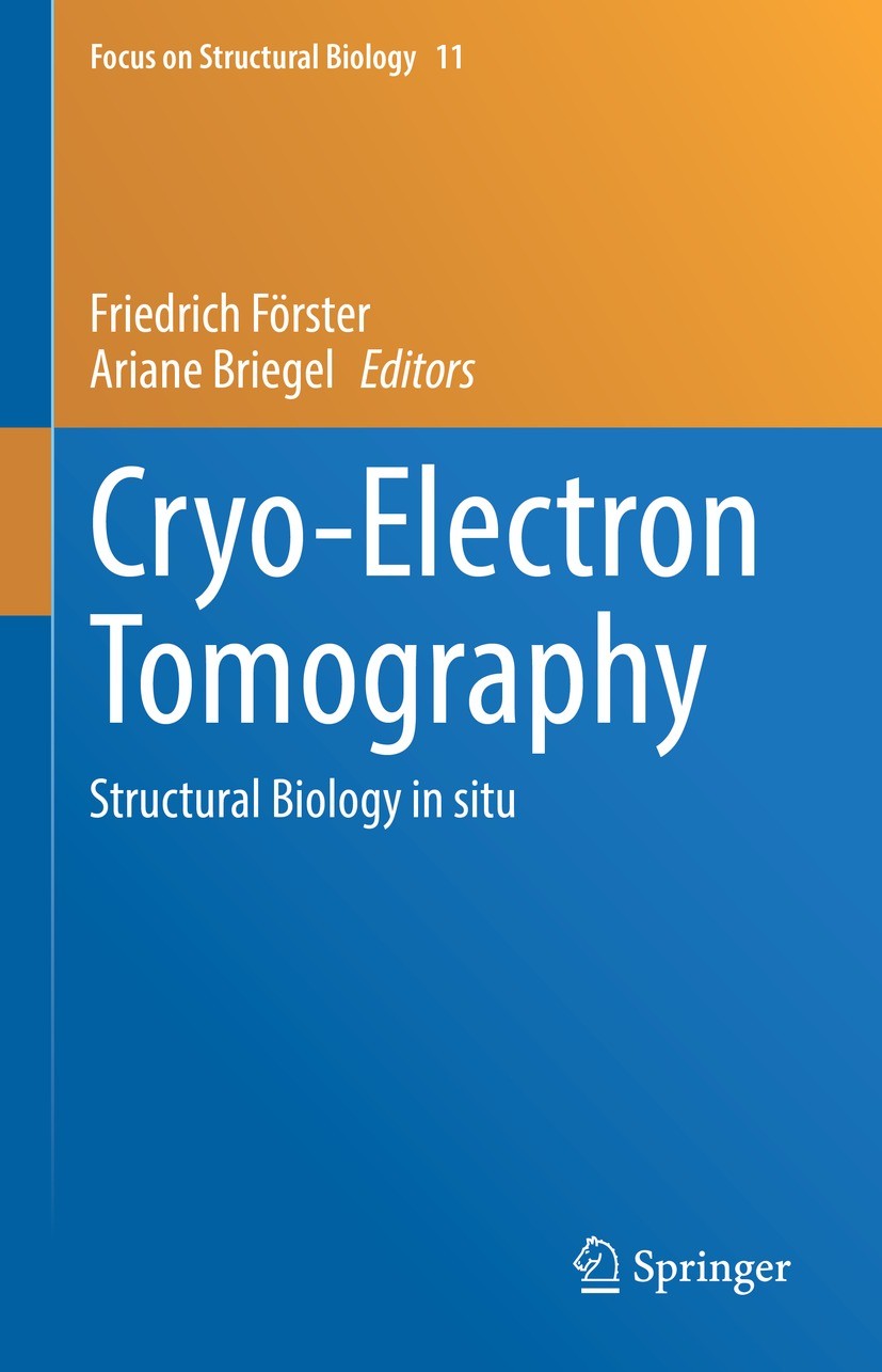
书目名称 | Cryo-Electron Tomography | 副标题 | Structural Biology i | 编辑 | Friedrich Förster,Ariane Briegel | 视频video | http://file.papertrans.cn/241/240489/240489.mp4 | 概述 | Comprehensive description of cryo-electron tomography.Addresses the underlying principles and all recent developments of cryo-ET.A valuable resource for scientists working in the field of structural b | 丛书名称 | Focus on Structural Biology | 图书封面 |  | 描述 | .This book presents key aspects and recent developments of cryogenic sample electron tomography (cryo-ET) methodology, authored by leading experts in the field. Understanding structure and function of biomolecules in the context of cells is a new frontier in cellular and structural biology. To facilitate such research, cryo-ET is a key method to visualize the molecules of life in their native settings. Cryo-ET enables the imaging of samples that are preserved in a near-native state, at (macro)-molecular resolution and in three dimensions. Thus, this technique is a unique tool to gain insights into how biomolecules collaborate in orchestrating fundamental biological processes, how mutations cause diseases, pathogens cause infections, and to develop novel therapeutics to treat such illnesses. This book provides a unique reference for the emerging field of cryo-ET.. The topics covered range from the fundamental principles of imaging to sample preparation, data analysis,and data sharing within the scientific community. It serves as a valuable resource for the next generation of structural biologists, making it suitable both for undergraduate students studying biochemistry, biophysics, | 出版日期 | Book 2024 | 关键词 | electron optics; phase contrast; tomography; tomographic reconstruction; Fourier-slice theorem; cryo-prep | 版次 | 1 | doi | https://doi.org/10.1007/978-3-031-51171-4 | isbn_softcover | 978-3-031-51173-8 | isbn_ebook | 978-3-031-51171-4Series ISSN 1571-4853 Series E-ISSN 2542-9566 | issn_series | 1571-4853 | copyright | The Editor(s) (if applicable) and The Author(s), under exclusive license to Springer Nature Switzerl |
1 |
Front Matter |
|
|
Abstract
|
2 |
,Fundamentals of Instrumentation and Electron Optics for Cryo-Electron Tomography, |
Juergen M. Plitzko,Daniel Bollschweiler |
|
Abstract
This chapter takes a close look at the hardware side of transmission electron microscopy and, by extension, its applications in cryo-electron tomography. Before we throw ourselves into the fray, we are briefly soaking in some information on notable development milestones of the past. I firmly believe there is value in appreciating the historical context of electron microscopy to gain a better understanding of the many advances that were necessary to get us to where we are today. In the chapter’s main part, we will then learn how to “make” electrons and how to make . of them in imaging techniques. We will deal with electron lenses and how an electron microscope is designed to create a space for the electron beam to shape and interact with our sample. We will discover the various ways in which these interactions take place and how, through the principle of phase contrast, a “micrograph” is eventually recorded in the image plane—by ever more sophisticated detectors. As we acknowledge that our sample fries all too quickly, we l also discuss low dose techniques and phase plates. Finally, we provide a short outlook towards exciting developments that may well push cryo-electron tomography
|
3 |
,Principles of Tomographic Reconstruction, |
Achilleas S. Frangakis |
|
Abstract
Imagine you need to look inside a living organism. You would have only two options: using invasive techniques or applying tomography. In this non-invasive imaging method, two-dimensional (2D) images of slices, or sections, within an organism are analyzed to determine the three-dimensional (3D) localization of internal objects. These sections can be so thin that the target objects are unobscured by the superposition of over- or underlying structures. The challenge of seeing internal objects that are free of superposition originated in medicine, where such 3D localization is necessary for accurate diagnosis and treatment.
|
4 |
,Cryogenic Preparations of Biological Specimens for Cryo-Electron Tomography, |
Edoardo D’Imprima,Herman K. H. Fung,Ievgeniia Zagoriy,Julia Mahamid |
|
Abstract
Cryo-electron tomography (cryo-ET) has become a method of choice for in-cell structural biology studies by integrating the non-invasive power of fluorescence light microscopy, artifact-free thinning of intact cells, and bridging to the atomic resolution achieved by single-particle cryo-electron microscopy (cryo-EM) of biochemically purified macromolecules. While the latter has reached a level of maturity and standardization for specimen preparation, cryo-ET needs to meet a multitude of requirements posed by its highly heterogenous sample types. Cryo-ET is compatible with samples prepared by both plunge-freezing and high-pressure freezing. Still, little is known about the vitrification process of complex and heterogenous cells during cryogenic specimen preparation. Thus, we first describe the physics of vitrification with emphasis on biological specimens prepared on EM-compatible supports. We then provide a comprehensive account of the current state-of-the-art cryo-ET sample preparation techniques for specimens ranging from in-vitro reconstitutions, single cells, to 3D cell cultures, and whole multicellular organisms. We point out specific considerations that facilitate a wide range
|
5 |
,Cryo-Focused Ion Beam Milling of Cells, |
Digvijay Singh,Elizabeth Villa |
|
Abstract
Cryo-electron tomography (cryo-ET) is a robust technique for bringing structural biology into the cellular context. However, transmission electron microscopes (TEM) used for cryo-ET are limited to samples up to 500 nm in thickness, preventing applicability of cryo-ET for most cells. As a result, cellular samples have to be thinned. Traditional EM fixation techniques help overcome this thickness limitation by resin-embedding samples that can be cut into thinner sections. Alternatively, Focused ion beam (FIB) milling is a well-established technique that nanomachines solid-state materials using the FIB without using chemical or mechanical methods. By adapting it to cryogenic conditions, delicate biological materials can be gently and precisely milled to produce thin slices suitable for cryo-ET. Vitrification preserves native structures of biomolecular networks and provides structural stability to frozen cells. Preparing samples using FIB milling of frozen-hydrated cells at cryogenic temperatures is known as cryo-FIB milling, which has emerged as one of the gold-standard techniques for cellular cryo-ET to generate molecular landscapes inside cells. In this chapter, we present a brief h
|
6 |
,In Situ Cryo-Electron Tomography and Advanced Micromanipulator Techniques, |
Sven Klumpe,Philipp S. Erdmann |
|
Abstract
cryo-electron tomography is undergoing a revolution as more challenging samples are moving into the center of attention. The complexity of the targeted structures is rapidly expanding and requires that preparation techniques evolve to keep up with the growing demand for larger specimens. In particular, organoids and tissue, but also small animals that can be vitrified in whole by high-pressure freezing, are targets of this new branch of in situ cryo-electron microscopy, which seeks to provide a “biopsy at the nanoscale”. With its recent coming of age, cryo-lift out is responding to this call. Novel hardware now makes this preparation technique commercially available to a broad audience without the need for significant prior knowledge or experience. Thanks to this, cryo-lift out is becoming increasingly popular and, through the concerted effort of the research community, more versatile and higher throughout. The following chapter provides a topical overview of this new and rapidly developing technique. We introduce the basic principles of cryo-lift out and briefly summarize the technological and biological results so far.
|
7 |
,Targeting, Localisation and Identification in Cryo-ET, |
Rainer Kaufmann,Kay Grünewald,Lindsay A. Baker |
|
Abstract
One of the main benefits of cryo-electron tomography (ET) is direct visualisation of the sample itself, without the need for stains or fixation. A consequence of this direct imaging is that every molecule in the sample contributes to the resulting tomogram, and for complex specimens such as cells or viruses, it can be challenging to identify a specific feature of interest. This chapter discusses two approaches to help guide collection and interpretation of data from cryo-ET: correlative light and electron microscopy (CLEM) and molecular tagging. These methods can be used independently or together and help cryo-ET practitioners identify areas for data collection and localise or identify specific molecules in their reconstructed tomograms.
|
8 |
,Automated Cryo Electron Tomography Data Collection, |
Wim J. H. Hagen |
|
Abstract
Electron tomography entails the three-dimensional reconstruction of an object from its projections, which are acquired using a transmission electron microscope (TEM). This chapter describes the process of collecting consecutive images of the tilted specimen efficiently in theory and practice. Firstly, the concept of a TEM and its main components are described prior to introducing the technical means to maintain an area of interest in the field of view during tilting. Then different tilt schemes are introduced as well as current approaches to maximize throughput and obtained signal per electron dose. A more detailed description of the TEM setup and different sample properties then allows a hands-on description of tilt series collection and its pitfalls. Lastly, technical innovations are discussed that may improve the optical quality of tiltseries in the coming years. In summary, this chapter provides the conceptual and practical framework for state-of-the-art data acquisition.
|
9 |
,Subtomogram Averaging, |
|
|
Abstract
|
10 |
,High-Resolution Tomography, Molecular Model Building and Visualization, |
Alberto Bartesaghi |
|
Abstract
A long sought-after goal in cellular biology is to be able to visualize macromolecular complexes inside cells at resolutions that are sufficient to characterize molecular level interactions. While cryo-electron tomography (ET) is uniquely positioned to address this challenge, numerous technical bottlenecks in data collection and image processing exist that have slowed down progress toward achieving this goal. This chapter describes the main challenges in high-resolution cryo-ET imaging and presents effective strategies to accelerate the collection of high-quality tilt-series, data-driven methods to efficiently extract high-resolution information from the raw data, strategies for tomogram segmentation and visualization, and techniques for atomic model building and integrative modeling of multi-component complexes imaged by cryo-ET. While challenges still remain in all these areas, this technology can already produce . atomic models of proteins in their native state, indicating that cryo-ET is on a trajectory to become an effective technique for visualizing cell interiors at unprecedented levels of detail.
|
11 |
,Beam-Induced Motion Mechanism and Correction for Improved Cryo-Electron Microscopy and Cryo-Electro |
Shawn Zheng,Axel Brilot,Yifan Cheng,David A. Agard |
|
Abstract
Beam induced motion (BIM) is a major cause of information loss in single-particle cryo-electron microscopy (cryo-EM) and cryo-electron tomography (cryo-ET). In this chapter we review the progress made in understanding the mechanism of BIM and methodologies developed to restore the information loss, in particular, in the context of cryo-ET. Observed data supports a doming model of BIM dominated by out-of-sample-plane motion and where heterogenous projections of local motions are observed. Sample tilting amplifies the apparent BIM as the out of plane motion becomes directly visible. This leads to more rapid motion at early doses as well as strong anisotropic motion in images recorded at high tilt angles. Based on the study of the BIM occurring within cryo-ET tilt series with very fine dose sampling, it was found that frozen hydrated samples, under repeated exposures, experience a “drumbeat” response, having both a repeated elastic component and a plastic deformation. The elastic deformation represents the portion that can be fully relaxed when the electron beam is turned off at the end of the exposure for each tilted image. By contrast, the plastic deformation decays exponentially wi
|
12 |
,Detection and Interpretation of Cellular Structures in Tomograms: Segmentation, Localization and Sp |
Antonio Martinez-Sanchez,Vladan Lučić |
|
Abstract
A wealth of information is present in cellular cryo-ET images, from the shape of large three-dimensional cellular structures to structural details of molecular complexes. However, extracting this information is particularly challenging for complexes that cannot be identified visually and are embedded in cellular environments comprising multitude of macromolecular species that lack an obvious ordering pattern. To overcome these challenges, a number of diverse computational methods were developed for the localization, molecular identification and tracing of cellular structures. Here we review diverse computational methods that were developed for segmentation of large subcellular structures, template-based and template-free localization of complexes, and analysis of their spatial organization. We cover a wide array of method complexity, from computer-assisted human-based visualizations to machine learning and advanced algorithms, and discuss challenges posed by the recent developments of deep learning methods. New approaches for localization of molecular complexes and spatial analysis are expected to provide precise molecular maps of diverse cellular compartments, elucidate the organi
|
13 |
,Electron Tomography of Cryo-Fixed and Resin-Embedded Samples, |
Alicia C. Borgeaud,Alejandro Melero,Lazar Ivanović,Wanda Kukulski |
|
Abstract
Room-temperature electron tomography of sections prepared from high-pressure frozen, freeze-substituted and resin-embedded cells is a powerful method to study intracellular architecture. The accessibility and versatility of this approach permit addressing many ultrastructural questions in cell and tissue biology. Such questions span different scales, from the localisation and organisation of protein assemblies to intricate cell remodeling processes during development. The robustness and high throughput of room-temperature electron tomography of resin samples facilitate identification and characterization of rare or transient cellular events as well as compilation of large datasets for quantitative analysis. Resistance to radiation damage enables dual-axis and montage tomography acquisitions for more isotropic resolution and for imaging large fields of view, respectively, thereby providing a wider context with equal detail. Electron tomography can also be performed on serial sections which allows entire cells or tissue volumes to be reconstructed at high resolution. Further, high precision correlative microscopy workflows can be implemented, permitting to combine information on mole
|
14 |
,After the Microscope: Long-Term Care of Electron Tomography Data, |
Catherine M. Oikonomou,Grant J. Jensen |
|
Abstract
Cryogenic electron tomography (cryo-ET) produces datasets tremendously rich in information, with significant value beyond their originating study. Openly sharing such data enables validation of research findings, sparks further biological discoveries, and fuels development of improved software tools for data processing. Data sharing is complicated, however, by the size of cryo-ET datasets. Here we synthesize our experience archiving and sharing nearly 20 years’ worth of cryo-ET data from our own research group to highlight general challenges and solutions for democratizing cryo-ET data and making it Findable, Accessible, Interoperable and Reusable long-term.
|
|
|