找回密码
 To register

QQ登录

只需一步,快速开始

扫一扫,访问微社区

Titlebook: CT and MRI of Skull Base Lesions; A Diagnostic Guide Igor Pronin,Valery Kornienko Book 2018 Springer International Publishing AG 2018 CT-Pe

[复制链接]
查看: 39733|回复: 59
发表于 2025-3-21 17:09:31 | 显示全部楼层 |阅读模式
书目名称CT and MRI of Skull Base Lesions
副标题A Diagnostic Guide
编辑Igor Pronin,Valery Kornienko
视频video
概述Equips readers with the knowledge required for accurate and timely diagnosis of skull base lesions.Presents appearances on both standard and new CT and MRI technologies, including CT perfusion.Offers
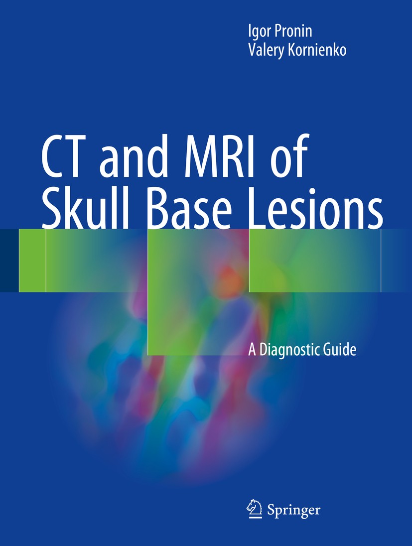
图书封面Titlebook: CT and MRI of Skull Base Lesions; A Diagnostic Guide Igor Pronin,Valery Kornienko Book 2018 Springer International Publishing AG 2018 CT-Pe
描述.This superbly illustrated book offers a comprehensive analysis of the diagnostic capabilities of CT and MRI in the skull base region with the aim of equipping readers with the knowledge required for accurate, timely diagnosis. The authors’ vast experience in the diagnosis of skull base lesions means that they are ideally placed to realize this goal, with the book’s contents being based on more than 10,000 histologically verified cases of frequent, uncommon, and rare diseases and disorders. In order to facilitate use, chapters are organized according to anatomic region. Readers will find clear guidance on complex diagnostic issues and ample coverage of appearances on both standard CT and MRI methods and newer technologies, including especially CT perfusion, susceptibility- and diffusion-weighted MRI (SWI and DWI), and MR spectroscopy. The book will be an ideal reference manual for neuroradiologists, neurosurgeons, neurologists, neuro-ophthalmologists, neuro-otolaryngologists, craniofacial surgeons, general radiologists, medical students, and other specialists with an interest in the subject..
出版日期Book 2018
关键词CT-Perfusion; Differential Diagnosis; CT-Perfusion; Magnetic Resonance Imaging; Computed Tomography; Diff
版次1
doihttps://doi.org/10.1007/978-3-319-65957-2
isbn_softcover978-3-319-88138-6
isbn_ebook978-3-319-65957-2
copyrightSpringer International Publishing AG 2018
The information of publication is updating

书目名称CT and MRI of Skull Base Lesions影响因子(影响力)


书目名称CT and MRI of Skull Base Lesions影响因子(影响力)学科排名


书目名称CT and MRI of Skull Base Lesions网络公开度


书目名称CT and MRI of Skull Base Lesions网络公开度学科排名


书目名称CT and MRI of Skull Base Lesions被引频次


书目名称CT and MRI of Skull Base Lesions被引频次学科排名


书目名称CT and MRI of Skull Base Lesions年度引用


书目名称CT and MRI of Skull Base Lesions年度引用学科排名


书目名称CT and MRI of Skull Base Lesions读者反馈


书目名称CT and MRI of Skull Base Lesions读者反馈学科排名


单选投票, 共有 1 人参与投票
 

0票 0.00%

Perfect with Aesthetics


1票 100.00%

Better Implies Difficulty


0票 0.00%

Good and Satisfactory


0票 0.00%

Adverse Performance


0票 0.00%

Disdainful Garbage

您所在的用户组没有投票权限
发表于 2025-3-21 22:00:56 | 显示全部楼层
发表于 2025-3-22 03:56:04 | 显示全部楼层
Benign Tumors as lesions of abnormal bone development (Paget’s disease, fibrous dysplasia, cementoossifying dysplasia, giant cell granuloma), benign tumors (exostosis, osteoma, osteochondroma, osteoid osteoma, osteoblastoma, ossifying fibroma, chondroma, and giant cell tumor), and malignant tumors, such as osteo
发表于 2025-3-22 05:34:25 | 显示全部楼层
发表于 2025-3-22 09:57:30 | 显示全部楼层
发表于 2025-3-22 15:22:11 | 显示全部楼层
发表于 2025-3-22 19:02:08 | 显示全部楼层
Sellar and Parasellar Tumorss) 10–15% of all intracranial tumors. These are mainly adult cases; these tumors are rarely seen in children, less than 6% of intracranial tumor in adolescents, rare in children (Osborn et al. 2016). The peak incidence is between 20 and 40 years. In rare cases, pituitary carcinoma may grow from the
发表于 2025-3-23 01:09:46 | 显示全部楼层
Book 2018adiologists, neurosurgeons, neurologists, neuro-ophthalmologists, neuro-otolaryngologists, craniofacial surgeons, general radiologists, medical students, and other specialists with an interest in the subject..
发表于 2025-3-23 02:09:27 | 显示全部楼层
Matthias Auchmann,Guillaume Urvoy-Kellerstic lesions, inflammatory changes in the sinuses of the skull, different types of polyps, which, growing into the ACF region, can mimic neoplasms, are often encountered in radiological practice. Imaging of most ACF and CFR lesions is primarily aimed at determining the expansion of the lesion rather
发表于 2025-3-23 06:29:12 | 显示全部楼层
Balaji Rengarajan,Gustavo de Vecianadermal movement of the face that take place in early period of embryogenesis (Shugar et al. 1980; Miro et al. 1998; Som et al. 2011). Chordomas don’t have a capsule and grow expansively with tumor “finger-like” outgrowths formation. If histologically tumors have cartilaginous inclusions, they are re
 关于派博传思  派博传思旗下网站  友情链接
派博传思介绍 公司地理位置 论文服务流程 影响因子官网 SITEMAP 大讲堂 北京大学 Oxford Uni. Harvard Uni.
发展历史沿革 期刊点评 投稿经验总结 SCIENCEGARD IMPACTFACTOR 派博系数 清华大学 Yale Uni. Stanford Uni.
|Archiver|手机版|小黑屋| 派博传思国际 ( 京公网安备110108008328) GMT+8, 2025-7-5 06:29
Copyright © 2001-2015 派博传思   京公网安备110108008328 版权所有 All rights reserved
快速回复 返回顶部 返回列表